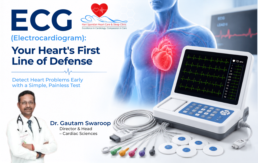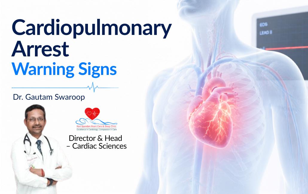
A Educational banner for Hari Spandan Heart Care & Sleep Clinic titled "Rhythms of the Rain: How the Monsoon Impacts Your Heart," featuring a portrait of Interventional Cardiologist Dr. Gautam Swaroop.

Rhythms of the Rain: How the Monsoon Impacts Your Heart

A Educational banner for Hari Spandan Heart Care & Sleep Clinic titled "Rhythms of the Rain: How the Monsoon Impacts Your Heart," featuring a portrait of Interventional Cardiologist Dr. Gautam Swaroop.

Quick Answer: Does the Monsoon Affect Your Heart?

Yes, it absolutely does. Specifically, the monsoon season introduces high humidity and extreme temperature drops. Furthermore, it brings a massive surge in dangerous viral infections. Consequently, these factors force your heart to work much harder.

Therefore, it struggles to regulate your core body temperature properly. Additionally, cold weather causes your blood vessels to constrict rapidly. Consequently, this raises your blood pressure dangerously high. Ultimately, you must prioritize extra cardiovascular care during the rain.

Introduction

The arrival of the monsoon is always a welcome relief. First, the cool breeze brings a much-needed summer break. However, your heart might be working in overdrive currently. Specifically, high humidity creates unique cardiovascular challenges for your body.

Furthermore, fluctuating temperatures stress your bodily systems immensely. At Harispandan Clinic, we see increased cardiac complaints daily during this season. Consequently, understanding these weather shifts remains absolutely critical. Therefore, you must protect your cardiovascular system proactively today.


Table of Contents


The Humidity Trap: Why Your Heart Works Harder

First, high humidity defines the rainy season completely. Specifically, thick moisture prevents your sweat from evaporating easily. Consequently, your core body temperature stays dangerously elevated. Therefore, your heart must pump much harder and faster.

Next, it pushes blood to your skin for rapid cooling. For healthy individuals, this acts as a minor workout. However, a weakened heart struggles with this added workload. Consequently, it triggers severe fatigue, breathlessness, or dangerous angina.

Temperature Fluctuations and Blood Pressure

The monsoon brings sudden, dramatic drops in temperature. Specifically, this happens during heavy downpours or late evenings. Consequently, your body gets cold very suddenly. Therefore, your blood vessels naturally narrow to conserve body heat.

Doctors call this dangerous biological process vasoconstriction. Furthermore, narrower vessels leave less space for blood to flow. Consequently, this instantly drives up your blood pressure. Ultimately, these sudden spikes significantly increase your heart attack risk.

The Threat of Monsoon Infections

The rainy season brings dangerous mosquito-borne illnesses rapidly. Furthermore, water-borne viral infections spread easily during this damp time. How does this specifically affect your heart?

  • Systemic Inflammation: First, your body fights severe infections aggressively. Consequently, it creates widespread internal inflammation. Therefore, this damages the delicate inner lining of your blood vessels. Ultimately, it makes arterial plaques highly unstable and causes clots.
  • Myocarditis: Next, certain viral infections attack the heart muscle directly. Consequently, this causes a dangerous condition called myocarditis. Therefore, the heart weakens significantly. Ultimately, this causes severe arrhythmias or even acute heart failure.
  • Fluid Imbalance: Finally, stomach bugs cause severe vomiting or diarrhea. Consequently, this leads to rapid, dangerous dehydration. Therefore, you lose critical electrolytes like potassium and sodium. Ultimately, you need these to maintain a normal heart rhythm.

The Dietary Trap: Cravings vs. Cardiac Health

Rainy weather brings intense cravings for deep-fried, salty foods. While comforting, these monsoon favorites remain highly dangerous. Specifically, they contain massive amounts of harmful trans fats. Consequently, these fats spike your bad cholesterol rapidly.

Therefore, they contribute heavily to dangerous arterial blockages. Furthermore, these popular snacks contain excess sodium. Consequently, sodium forces your body to retain extra water. Ultimately, this drives up your blood pressure instantly.

The Sedentary Slump

For many, the monsoon ends their daily morning walk. Specifically, mud and unpredictable downpours make outdoor exercise highly difficult. Consequently, stopping your exercise routine causes cardiovascular problems immediately. Therefore, your resting heart rate rises rapidly.

Furthermore, your blood pressure creeps up silently over time. Next, your blood sugar levels become highly unstable. Ultimately, this sedentary behavior destroys months of hard cardiovascular conditioning.

5 Tips for a Heart-Healthy Monsoon

At Harispandan Clinic, we highly recommend these actionable steps. Specifically, these daily tips protect your heart during the rain.

  • Move Indoors: First, do not let the rain stop your routine. Instead, switch to indoor activities like yoga immediately. Furthermore, try spot jogging or stair climbing at home safely.
  • Smart Snacking: Next, satisfy your cravings with heart-healthy alternatives. Specifically, try roasted corn with lemon instead of heavy salt. Furthermore, eat baked sweet potatoes or unsalted mixed nuts.
  • Stay Hydrated: People often forget to drink water during cooler weather. Consequently, dangerous dehydration thickens your blood significantly. Therefore, your heart must work much harder. Ultimately, keep sipping warm water or herbal teas daily.
  • Boost Your Immunity: First, protect yourself from dangerous viral infections proactively. Specifically, eat a diet rich in Vitamin C. Furthermore, maintain strict hand hygiene every single day. Therefore, ensure your surroundings remain completely free of stagnant water.
  • Monitor Your Blood Pressure: Specifically, hypertensive patients must remain highly vigilant. Therefore, check your blood pressure at home more frequently. Consequently, you can catch weather-induced spikes very early.

Frequently Asked Questions

  • Is it safe to drink tea during the monsoon?
    Yes, moderate consumption remains generally safe. However, excessive caffeine increases your heart rate dangerously. Therefore, opt for green tea or ginger tea instead.
  • Can a cold shower cause a heart attack?
    Yes, sudden cold exposure acts as a dangerous trigger. Specifically, it causes a massive spike in blood pressure. Therefore, always dry off quickly and take warm showers.
  • Why do I feel breathless during the monsoon?
    First, high humidity makes the air feel much heavier. Consequently, your heart works harder to cool the body. Therefore, fluid can back up into your lungs easily. Ultimately, this causes severe breathlessness.

Key Takeaways

  • First, high humidity forces the heart to pump harder.
  • Consequently, this strains compromised cardiovascular systems severely.
  • Next, sudden temperature drops cause blood vessels to constrict rapidly.
  • Therefore, this leads to highly dangerous blood pressure spikes.
  • Furthermore, viral infections cause dangerous systemic inflammation.
  • Finally, always avoid deep-fried, salty monsoon snacks entirely.

Protect Your Heart This Season

The monsoon should remain a wonderful time for rejuvenation. However, you must monitor unusual symptoms like chest heaviness carefully. At Harispandan Clinic, we provide comprehensive cardiac evaluations daily. Consequently, we ensure your heart remains strong and healthy.

Therefore, book your monsoon health screening with us today. Ultimately, you can enjoy the beautiful rhythms of the rain peacefully.

Medical Disclaimer
Please note, this article provides educational information only. It never replaces professional medical advice, diagnosis, or treatment. Always follow the specific advice of your treating cardiologist. Furthermore, seek immediate emergency medical attention for severe chest pain, shortness of breath, or sudden fainting.

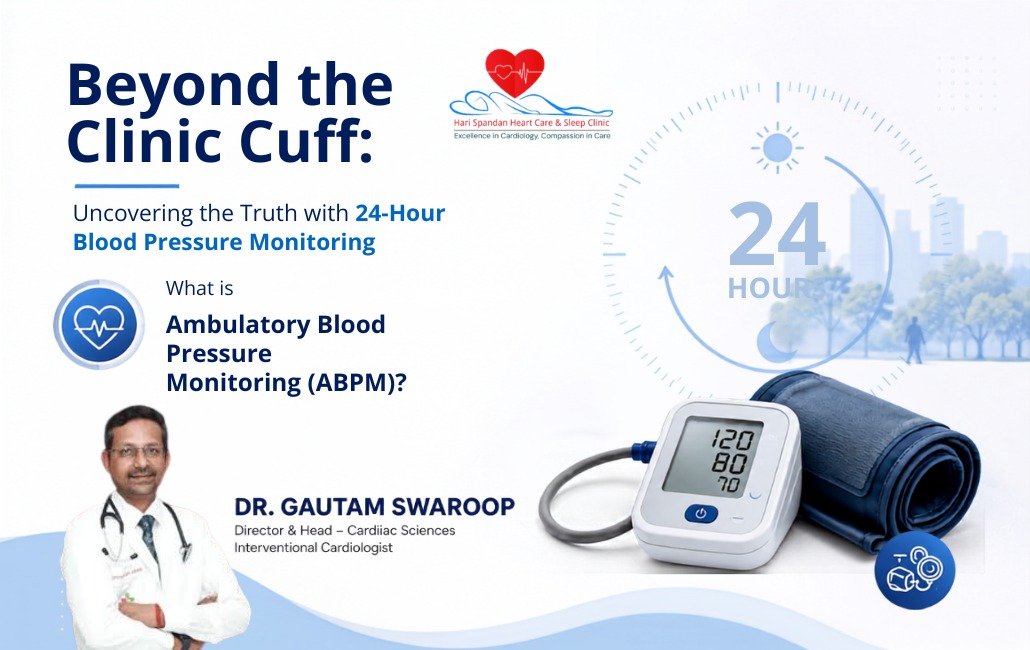
Professional minimal healthcare banner illustrating 24-hour Ambulatory Blood Pressure Monitoring (ABPM). A digital blood pressure monitor with cuff is placed beside a subtle 24-hour clock graphic, representing continuous blood pressure tracking throughout the day and night. The design features a clean white and blue medical theme with abstract healthcare elements, promoting awareness of ABPM in a modern clinical setting.

Beyond the Clinic Cuff: Uncovering the Truth with 24-Hour Blood Pressure Monitoring

Professional minimal healthcare banner illustrating 24-hour Ambulatory Blood Pressure Monitoring (ABPM). A digital blood pressure monitor with cuff is placed beside a subtle 24-hour clock graphic, representing continuous blood pressure tracking throughout the day and night. The design features a clean white and blue medical theme with abstract healthcare elements, promoting awareness of ABPM in a modern clinical setting.

Quick Answer: What is Ambulatory Blood Pressure Monitoring (ABPM)?

Ambulatory Blood Pressure Monitoring (ABPM) is a highly effective, non-invasive test. Specifically, it records your blood pressure automatically over a full 24-hour period. You simply wear a small digital cuff while going about your normal daily life.

Consequently, the device takes accurate readings every 20 to 30 minutes during the day. Furthermore, it continues checking every 30 to 60 minutes while you sleep. Therefore, it provides a comprehensive picture of your cardiovascular health. Ultimately, a single clinic reading simply cannot capture this vital daily data.

Introduction

Imagine taking your car to a mechanic for a strange highway noise. However, when the mechanic idles the engine in the garage, it sounds perfectly fine. Diagnosing hypertension often presents a remarkably similar medical challenge.

Specifically, your blood pressure is never a static number. It constantly fluctuates based on your daily activity, stress levels, and sleep. When you visit a clinic, the doctor gets just a single snapshot at that exact moment. But what happens during the other 23 hours of your day?

At Harispandan Clinic, we rely heavily on Ambulatory Blood Pressure Monitoring to solve this mystery. By shifting from a single snapshot to a continuous 24-hour movie, we easily diagnose hidden heart risks. Consequently, we prevent unnecessary medications and tailor your cardiovascular treatment with pinpoint precision.


Table of Contents


Why a Single Clinic Reading Isn’t Always Enough

Relying purely on isolated in-office readings often leads to frustrating misdiagnoses. Specifically, two very common psychological phenomena cause these medical errors.

1. White Coat Hypertension

Do you feel nervous at the doctor’s office? You are definitely not alone. The intense stress of traffic, waiting rooms, and medical exams temporarily spikes your blood pressure.

Doctors call this widespread phenomenon White Coat Hypertension. Without ABPM, a doctor might misdiagnose you with chronic high blood pressure. Consequently, they might prescribe heavy medication you do not actually need. Therefore, your pressure could drop dangerously low when you finally relax at home.

2. Masked Hypertension

Conversely, this represents the dangerous opposite of the White Coat effect. Specifically, a patient’s numbers read perfectly normal in the calm, quiet clinic environment. However, they return to stressful jobs and poor dietary habits quickly.

Consequently, their pressure spikes to highly dangerous levels during normal routines. Therefore, a 24-hour monitor successfully strips away this deceptive mask. Ultimately, it reveals your true, underlying cardiovascular risk clearly.

What ABPM Catches That a Regular Check Misses

Beyond filtering out simple clinic-induced anxiety, 24-hour monitoring provides absolutely critical data. Specifically, it shows exactly how your heart behaves when you stop paying attention to it.

Crucial Medical Insight: Nighttime “Dipping”

In a perfectly healthy cardiovascular system, your numbers naturally drop while you sleep. Doctors expect a healthy nighttime dip of roughly 10% to 20%. However, if your numbers fail to dip, it signals a major red flag.

Furthermore, if your numbers actually rise during sleep, the danger increases significantly. Consequently, this non-dipping pattern strongly links to an increased risk of stroke and heart attack. Standard clinic readings completely miss this severe nocturnal danger.

Additionally, ABPM remains the absolute gold standard for evaluating medication efficacy. If you take blood pressure medication, the monitor reveals if the drug wears off too early. Therefore, doctors can adjust your exact dosage to cover dangerous early-morning spikes.

Standard BP Check vs. 24-Hour ABPM

Feature In-Clinic Blood Pressure Check Ambulatory Blood Pressure Monitoring (ABPM)
Duration of Test 1 to 5 minutes 24 continuous hours
Number of Readings Usually 1 to 3 readings 50 to 70+ automated readings
Patient Environment Clinical, often stressful setting Natural daily routine (home, work, sleep)
Nighttime Data Absolutely None Continuous, crucial nocturnal tracking
Best Used For Routine screening, checking immediate vitals Diagnosing white coat hypertension and evaluating drug efficacy

Who Needs an ABPM Test?

Your doctor at Harispandan Clinic may highly recommend 24-hour monitoring if you fit these clinical profiles:

  • You consistently show borderline high readings during routine clinic visits.
  • Your clinic readings remain high, but your home machine readings are completely normal.
  • You experience frequent fainting, dizziness, or lightheadedness unexpectedly.
  • Your numbers refuse to drop despite taking multiple prescribed medications daily.
  • You suffer from sleep apnea, kidney disease, or advanced diabetes currently.

What to Expect During Your Test at Harispandan Clinic

Undergoing an ABPM test is incredibly simple, painless, and completely non-invasive. Here is exactly how the process works at our dedicated clinic:

  • Fitting the Device: First, a technician wraps a standard cuff around your upper arm tightly. Next, they connect it to a lightweight digital monitor worn on a belt.
  • The 24-Hour Routine: You leave the clinic and simply go about your normal day. You must work, eat, relax, and sleep exactly as you normally would.
  • Automatic Readings: The cuff automatically inflates every 20-30 minutes during the day. Furthermore, it inflates every 30-60 minutes overnight.
  • Keeping a Diary: We ask you to briefly jot down your current activities when the cuff inflates. Consequently, you must note any sudden symptoms like dizziness.
  • Reviewing the Data: Finally, you return the device after 24 hours. Our cardiology specialists download the vast data and provide a highly accurate diagnosis.

Frequently Asked Questions

  • Does the inflating cuff hurt?
    No, it feels exactly like a normal manual check. Specifically, it squeezes your arm tightly for a few seconds and then releases completely.
  • Can I sleep with the monitor on?
    Yes, you absolutely must. While the initial inflations might briefly wake light sleepers, most patients adapt quickly. Ultimately, the sleep data is the most valuable part of the entire test.
  • Can I take a shower or exercise while wearing it?
    No, you cannot shower or swim while wearing the monitor. Specifically, the electronic device cannot get wet. Furthermore, we strongly recommend avoiding heavy, vigorous exercise during the testing period. Consequently, excessive arm movement triggers annoying error readings.
  • What should I do when I feel the cuff inflating?
    If you are walking, you must simply stop moving immediately. Next, keep your arm completely relaxed and straight at your side. Ultimately, this ensures the machine grabs an accurate measurement on the first attempt.

Key Takeaways

  • A single clinic reading often misleads doctors due to extreme patient stress.
  • ABPM records your numbers over 50 times to provide a true cardiovascular picture.
  • It remains the only accurate way to detect dangerous non-dipping nighttime levels.
  • The test is completely painless and worn during your normal daily routine.
  • Ultimately, it helps doctors prescribe the exact right amount of medication needed.

Schedule Your Assessment Today

Do not leave your critical heart health to simple guesswork or isolated clinic readings. If you struggle to control your blood pressure, it is time to look beyond the basic cuff.

At Harispandan Heart Care & Sleep Clinic, we use state-of-the-art Ambulatory Blood Pressure Monitoring technology daily. Consequently, we provide you with the most accurate cardiovascular diagnosis possible. Contact our dedicated cardiology team today to schedule your immediate ABPM fitting.

Medical Disclaimer
Please note, this article provides educational information only. It never replaces professional medical advice, diagnosis, or treatment. Always follow the specific advice of your treating cardiologist. Furthermore, seek immediate emergency medical attention for severe chest pain, shortness of breath, or sudden fainting.

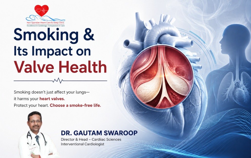
An educational medical poster from Hari Spandan Heart Care & Sleep Clinic titled "Smoking & Its Impact on Valve Health." Below a decorative heartbeat line, the text reads, "Smoking doesn't just affect your lungs— it harms your heart valves. Protect your heart. Choose a smoke-free life." On the bottom left is a portrait of Dr. Gautam Swaroop, Director & Head of Cardiac Sciences and Interventional Cardiologist. The right side features a large, detailed 3D illustration of a heart, with a circular magnified inset showing a detailed, colorful view of the inner heart valves. The background includes stylized wisps of smoke and a blue silhouette of a human torso highlighting the lungs and heart.

Up in Smoke: How Smoking Secretly Damages Your Heart Valves

An educational medical poster from Hari Spandan Heart Care & Sleep Clinic titled "Smoking & Its Impact on Valve Health." Below a decorative heartbeat line, the text reads, "Smoking doesn't just affect your lungs— it harms your heart valves. Protect your heart. Choose a smoke-free life." On the bottom left is a portrait of Dr. Gautam Swaroop, Director & Head of Cardiac Sciences and Interventional Cardiologist. The right side features a large, detailed 3D illustration of a heart, with a circular magnified inset showing a detailed, colorful view of the inner heart valves. The background includes stylized wisps of smoke and a blue silhouette of a human torso highlighting the lungs and heart.

Quick Answer: How Does Smoking Affect Heart Valves?

Smoking severely damages your heart valves rapidly. Specifically, it accelerates dangerous calcium buildup and raises your blood pressure. Furthermore, toxic chemicals destroy the delicate inner lining of the valves.

Consequently, they become stiff and narrowed, causing valve stenosis. Alternatively, they become weak and leaky, causing valve regurgitation. Therefore, your heart works dangerously hard. Ultimately, this leads to a massive risk of heart failure.

Introduction

People usually associate smoking with lung cancer and clogged arteries. However, a hidden mechanical danger occurs with every single puff. Specifically, smoking causes slow, irreversible damage to your heart valves.

Your heart relies on four delicate valves to direct blood flow. Consequently, they act as vital one-way doors. But, toxic chemicals break down their structural integrity quickly. At Harispandan Clinic, we see these devastating cardiovascular consequences daily.

Therefore, understanding this habit is crucial for your long-term health. Below, we explain exactly how smoking destroys valve tissue.


Table of Contents


The Role of Your Heart Valves

Your heart possesses four distinct valves. Specifically, these are the aortic, mitral, tricuspid, and pulmonary valves. First, they open completely to let blood flow forward. Next, they snap shut tightly to prevent dangerous backward flow.

Therefore, these valves must remain perfectly flexible and strong. However, damaged valves force your heart to pump much harder. Consequently, this constant strain leads to exhaustion and heart failure.

3 Ways Smoking Directly Destroys Valve Health

1. Accelerating Valve Calcification (Aortic Stenosis)

First, smoking drastically impacts your aortic valve. Specifically, tobacco chemicals trigger a massive inflammatory response in your bloodstream. Furthermore, this inflammation accelerates calcium deposits on the valve leaflets.

Consequently, the flexible valve becomes rigid, thick, and severely narrowed. Doctors call this dangerous condition Aortic Stenosis. Therefore, your heart must squeeze with immense force. Ultimately, it struggles to push blood through this tiny opening.

2. High Blood Pressure and Valve Strain (Regurgitation)

Next, nicotine acts as a powerful stimulant in your body. Specifically, it constricts your blood vessels and raises your blood pressure. Consequently, high pressure forces blood to slam against closed heart valves.

Over time, this intense pressure stretches and weakens the delicate tissue. Therefore, the valves simply cannot close properly anymore. Ultimately, this creates a leaky valve, known as valve regurgitation. Blood then spills backwards dangerously into your heart chambers.

3. Endothelial Destruction

Finally, smoking actively destroys your protective cell lining. Specifically, doctors call this delicate inner layer the endothelium. Furthermore, carbon monoxide and free radicals are highly toxic here.

Consequently, smoking literally strips away this vital protective layer. Therefore, your heart valves become highly susceptible to tearing. Ultimately, this damage promotes severe scarring and dangerous fatty plaques.

The Hidden Danger: Smoking, Gums, and Heart Infections

Interestingly, a strong link exists between smoking and oral health. First, smoking severely compromises your overall immune system. Consequently, it acts as a leading cause of severe gum disease.

Therefore, dangerous bacteria easily enter your bloodstream through infected gums. Furthermore, smoking has already damaged your valve surfaces. Thus, traveling bacteria rapidly latch onto the scarred tissue. Ultimately, this causes a life-threatening infection called Infective Endocarditis.

Healthy Valve vs. Smoker’s Valve

Feature Healthy Heart Valve Smoker’s Heart Valve
Tissue Flexibility Soft, thin, and highly flexible. Stiff, thick, and rigid.
Calcium Deposits None to minimal. Heavy, accelerated calcification.
Closure Quality Seals tightly, preventing backflow. May leak due to stretching and scarring.
Endothelium (Lining) Smooth and completely protective. Damaged, inflamed, and sticky.
Infection Risk Low risk of bacterial attachment. High risk of bacteria latching onto scars.

Can Quitting Reverse Valve Damage?

We hear this question frequently at Harispandan Clinic. Unfortunately, quitting smoking cannot reverse existing structural damage. Specifically, it will not magically dissolve heavy calcium deposits. Furthermore, it cannot repair a torn valve leaflet.

Consequently, severe cases absolutely require surgical repair or replacement. However, quitting smoking remains incredibly important for your survival. First, stopping tobacco halts the rapid acceleration of the damage. Next, it drastically lowers your blood pressure instantly.

Therefore, it reduces the massive strain on your healthy tissue. Ultimately, quitting significantly improves your surgical recovery chances.

Warning Signs of Heart Valve Disease

Heart valve disease often develops silently over decades of smoking. Therefore, you must watch for these critical red flags:

  • Unexplained shortness of breath during light physical activity.
  • A feeling of heavy pressure during exercise.
  • Severe heart palpitations causing a racing or fluttering heartbeat.
  • Noticeable swelling accumulating in your ankles, feet, or abdomen.
  • Frequent dizzy spells, lightheadedness, or sudden fainting episodes.
  • A distinctive heart murmur detected during a stethoscope exam.

Frequently Asked Questions

  • Does vaping also damage heart valves?
    Yes, absolutely. Vaping does not produce tar. However, e-cigarettes still contain dangerous nicotine. Consequently, this raises your blood pressure and heart rate. Therefore, vaping puts severe mechanical strain on your heart valves.
  • Are women who smoke at a higher risk for valve disease?
    Yes, they are. Studies show female smokers face significantly higher cardiovascular risks. Consequently, they experience more severe valve complications than male smokers.
  • How is heart valve disease diagnosed?
    At Harispandan Clinic, we primarily use an Echocardiogram. Specifically, this painless ultrasound lets us see your valves moving. Therefore, we can detect blood flow issues and calcium buildup easily.
  • Can secondhand smoke affect my heart valves?
    Yes, it certainly can. Prolonged exposure severely damages your endothelial lining. Consequently, secondhand smoke increases your risk for valve disease and heart failure.

Key Takeaways

  • Smoking directly attacks the structural integrity of your mechanical valves.
  • Tobacco chemicals accelerate calcium buildup, causing dangerous Aortic Stenosis.
  • Nicotine induces high blood pressure and causes valve regurgitation.
  • Damaged linings and gum disease increase your risk for Endocarditis.
  • Quitting smoking immediately stops the rapid progression of valve damage.

Protect Your Heart with Harispandan Clinic

Heart valve disease is progressive and worsens over time. This is especially true if you continue to smoke. Therefore, early detection remains the key to preventing irreversible damage. Consequently, we can explore non-surgical management options together.

Never wait for a massive medical emergency to strike. Harispandan Clinic utilizes state-of-the-art diagnostic technology for precise evaluations. Specifically, our advanced echocardiography maps your valve health perfectly. Take the first crucial step toward securing your cardiovascular future today. Ultimately, contact our clinic to schedule your comprehensive heart health screening.

Medical Disclaimer
Please note, this article provides educational information only. It never replaces professional medical advice, diagnosis, or treatment. Always follow the specific advice of your treating cardiologist. Furthermore, seek immediate emergency medical attention for severe chest pain, shortness of breath, or sudden fainting.

Professional Banner for Hari Spandan Heart Care & Sleep Clinic featuring Dr. Gautam Swaroop, Interventional Cardiologist, with a Holter monitor device and the headline 'Beyond the Snapshot: What a Holter Monitor Catches That a Regular EKG Misses.' The image highlights continuous heart rhythm monitoring to detect irregular heart conditions that may not appear on a standard ECG.

Beyond the Snapshot: What a Holter Monitor Catches That a Regular EKG Misses

Professional Banner for Hari Spandan Heart Care & Sleep Clinic featuring Dr. Gautam Swaroop, Interventional Cardiologist, with a Holter monitor device and the headline 'Beyond the Snapshot: What a Holter Monitor Catches That a Regular EKG Misses.' The image highlights continuous heart rhythm monitoring to detect irregular heart conditions that may not appear on a standard ECG.

Quick Answer: What is the Difference Between an EKG and a Holter Monitor?

An EKG (Electrocardiogram) is a quick, 10-second snapshot of your heart’s electrical activity taken in a clinic. A Holter Monitor is a wearable, continuous ECG device that records your heart’s activity for 24 to 48 hours.

While an EKG detects constant or immediate heart issues, a Holter monitor is essential for catching intermittent problems like sudden palpitations, hidden arrhythmias, or dizzy spells that happen during your normal daily routine.

Introduction

It is a frustratingly common scenario: You are sitting at home or working out when suddenly, your heart starts racing, fluttering, or skipping a beat. You feel dizzy and anxious. But by the time you schedule an appointment and sit in the cardiologist’s office for an Electrocardiogram (EKG), your heart is beating perfectly normally. The test comes back clear, yet you know exactly what you felt.

Are you imagining things? Absolutely not.

At Harispandan Clinic, we frequently see patients who experience this exact frustration. The heart is a dynamic organ, and its electrical system can glitch sporadically. Relying solely on a standard EKG is like looking at a single photograph of a busy intersection and trying to understand the traffic flow for the entire day.

To truly understand what is happening inside your chest, we often need to switch from a “photograph” to a “video camera.” That is exactly where the Holter Monitor comes in.


Table of Contents


The Standard EKG: The 10-Second Snapshot

An Electrocardiogram (EKG or ECG) is the foundation of cardiac diagnostics. It is painless, non-invasive, and takes only a few minutes. Electrodes are placed on your chest and limbs to record the electrical signals that make your heart beat.

What an EKG does well:

  • Detects a heart attack in progress or evidence of a past heart attack.
  • Identifies sustained arrhythmias (like atrial fibrillation) that are happening at the exact moment of the test.
  • Checks the basic structural electrical pathways of the heart.

The Limitation:

An EKG records roughly 10 to 15 seconds of heartbeats. If your heart happens to be behaving normally during that brief window, the EKG will show a clean bill of health—completely missing the abnormal rhythms that happen while you are sleeping, exercising, or feeling stressed.

Enter the Holter Monitor: The Continuous “CCTV”

A Holter monitor is essentially a portable, miniaturized EKG machine. It consists of a small recording device (roughly the size of a deck of cards) attached to electrodes on your chest. You wear it continuously as you go about your normal life—typically for 24 to 48 hours, though modern variations can be worn for up to two weeks.

Because it records continuously, it captures roughly 100,000 heartbeats in a single day, compared to the mere 10 or 15 beats captured by a standard clinic EKG.

What a Holter Monitor Catches That an EKG Misses

1. Intermittent Arrhythmias

An arrhythmia is an irregular heartbeat. It can beat too fast (tachycardia), too slow (bradycardia), or erratically. Many arrhythmias are paroxysmal, meaning they come and go suddenly. A Holter monitor acts as a 24-hour trap, capturing these fleeting electrical misfires the moment they happen, whether you are at your desk or out for a walk.

2. Symptom-to-Heartbeat Correlation

When you wear a Holter monitor from Harispandan Clinic, we ask you to keep a symptom diary. If you feel a sudden flutter at 2:15 PM, you write it down. Our specialists then cross-reference your diary with the monitor’s data at exactly 2:15 PM. This proves whether your symptoms are actually caused by a cardiac event or if they are related to something else, like anxiety or gastric reflux.

3. Silent Ischemia

Ischemia occurs when the heart muscle does not get enough oxygen-rich blood, often due to blocked arteries. While this usually causes chest pain (angina), it can sometimes happen without any symptoms at all—a condition known as silent ischemia. A standard EKG will miss this unless it happens in the clinic. A Holter monitor can detect the distinct electrical changes that signify blood flow restriction during your daily physical activities.

4. Sleep-Related Cardiac Events

Your heart rate naturally dips while you sleep. However, for some individuals—especially those with obstructive sleep apnea—breathing pauses can trigger dangerous drops in heart rate or sudden, alarming spikes. A standard daytime EKG cannot evaluate your heart’s behavior during the vulnerable hours of sleep.

Comparison: EKG vs. Holter Monitor

Feature Standard EKG (ECG) Holter Monitor
Duration of Test 10 to 15 seconds 24 to 48 hours (sometimes longer)
Setting Doctor’s office or hospital bed Worn at home, work, and during sleep
Best Used For Immediate emergencies, baseline heart health Unexplained fainting, intermittent palpitations
Patient Activity Lying perfectly still Normal daily routine
Number of Heartbeats Recorded ~10 to 15 ~100,000+ per day

Who Needs a Holter Monitor?

The specialists at Harispandan Clinic may recommend a Holter monitor if you experience any of the following unexplained symptoms:

  • Palpitations: A feeling that your heart is skipping beats, fluttering, or pounding hard.
  • Unexplained Syncope: Fainting or sudden bouts of extreme dizziness.
  • Shortness of Breath: Especially if it occurs suddenly without physical exertion.
  • Post-Treatment Monitoring: To ensure that newly prescribed anti-arrhythmic medications or a recently implanted pacemaker are working correctly.

What to Expect at Harispandan Clinic

If you require a Holter monitor, the process at our clinic is seamless and designed for your comfort.

  • Placement: A technician will clean small areas on your chest and attach sticky electrode patches.
  • Connection: Wires connect the electrodes to a small recording device, which you can clip to your belt or wear around your neck.
  • Your Routine: You will leave the clinic and go about your normal day. You can work, walk, and sleep normally. (Note: Most standard monitors cannot get wet, so you will need to avoid showering or swimming during the 24-hour testing period).
  • Logging: You will be given a small diary to log your activities and any symptoms you feel.
  • Review: Once you return the device, our cardiologists use advanced software to analyze the massive amount of data, cross-referencing it with your diary to formulate an accurate diagnosis.

Frequently Asked Questions

  1. Can I use my smartphone or smartwatch instead of a Holter monitor?
    While smartwatches with ECG features are fantastic for early detection and have revolutionized personal health tracking, they typically only record a single lead (one electrical view of the heart) and only when you actively trigger them. A Holter monitor records multiple leads continuously, providing the clinical-grade data a cardiologist needs to make a definitive diagnosis.
  2. Does wearing a Holter monitor hurt?
    Not at all. The test is entirely non-invasive. You might experience a mild, temporary skin irritation from the adhesive on the electrode patches, but there is no pain or electrical shock involved.
  3. Will the monitor interfere with my daily life?
    The devices are small and discreet under clothing. Aside from keeping the device dry and avoiding high-voltage electrical fields (like MRI machines), you are encouraged to stick to your normal routine so the monitor can capture a true reflection of your day.

Key Takeaways

  • A standard EKG is excellent for immediate diagnosis but only captures a 10-second snapshot of your heart’s electrical activity.
  • A Holter monitor is a 24-to-48-hour continuous recording that acts like a CCTV camera for your heart.
  • Holter monitors are crucial for catching intermittent palpitations, fainting spells, and sleep-related cardiac events that an EKG frequently misses.
  • Keeping an accurate symptom diary while wearing the monitor is vital for a precise diagnosis.

Schedule Your Cardiac Assessment

Heart symptoms can be anxiety-inducing, especially when initial tests come back normal while the symptoms persist. You know your body best. If you are experiencing unexplained palpitations, dizziness, or chest flutters, do not settle for a mere snapshot.

At Harispandan Clinic, we utilize advanced continuous monitoring technology to uncover the hidden rhythms of your heart. Reach out to our cardiology team today to schedule an evaluation and get the answers—and peace of mind—you deserve.

Medical Disclaimer
This article is intended for educational purposes only and should not replace professional medical consultation. If you are experiencing severe chest pain, shortness of breath, or prolonged fainting, seek emergency medical attention immediately.

A patient getting their blood pressure checked at Harispandan Clinic to screen for hypertension and prevent heart disease.

Hypertension (High Blood Pressure): Causes, Symptoms, Risks, Diagnosis, Treatment & Prevention

A patient getting their blood pressure checked at Harispandan Clinic to screen for hypertension and prevent heart disease.

Quick Answer: What Is Hypertension?

Hypertension, commonly known as high blood pressure, is a medical condition in which the pressure of blood against the walls of the arteries remains consistently higher than normal.

Over time, uncontrolled high blood pressure can damage the heart, blood vessels, kidneys, brain, and other vital organs. Because hypertension often develops without noticeable symptoms, many people may have the condition for years without knowing it. For this reason, hypertension is often called a “silent killer.”

Regular blood pressure monitoring, timely diagnosis, lifestyle modifications, and appropriate medical treatment can help control hypertension and significantly reduce the risk of serious complications such as heart attack, stroke, heart failure, and kidney disease.

Introduction

High blood pressure is one of the most common health conditions affecting adults worldwide. However, despite being extremely common, hypertension is frequently ignored because it may not cause obvious symptoms during its early stages. You may feel completely healthy while your blood pressure remains elevated.

Meanwhile, the increased pressure can gradually damage your arteries and force your heart to work harder to pump blood throughout your body. Over time, uncontrolled hypertension can increase the risk of coronary artery disease, heart attack, stroke, heart failure, kidney damage, and vision problems.

As cardiologists often explain to patients, blood pressure is similar to the pressure of water flowing through a pipe. When the pressure remains excessively high for a long period, it can gradually damage the walls of the pipe. Similarly, persistently high blood pressure can damage your blood vessels and vital organs.

Perhaps you have recently received a high blood pressure reading. Alternatively, you may have diabetes, high cholesterol, obesity, or a family history of hypertension. Or, you may simply want to understand how to protect your cardiovascular health. Regardless of the reason, understanding hypertension is an important step toward preventing serious health complications. This comprehensive guide explains what hypertension is, its causes, symptoms, risk factors, diagnosis, treatment options, complications, and preventive measures.

Table of Contents

  1. What Is Hypertension?
  2. Understanding Blood Pressure Readings
  3. What Causes Hypertension?
  4. Types of Hypertension
  5. What Are the Symptoms of High Blood Pressure?
  6. Risk Factors for Hypertension
  7. How Is Hypertension Diagnosed?
  8. What Tests May Be Recommended?
  9. Why Is Uncontrolled Hypertension Dangerous?
  10. Complications of High Blood Pressure
  11. How Is Hypertension Treated?
  12. Lifestyle Changes to Control Blood Pressure
  13. Medications Used for Hypertension
  14. How to Monitor Blood Pressure at Home
  15. When Should You See a Cardiologist?
  16. Frequently Asked Questions
  17. Key Takeaways
  18. Conclusion

What Is Hypertension?

Medical Definition

Hypertension is a medical condition in which the pressure exerted by circulating blood against the walls of the arteries remains consistently elevated.

Blood pressure naturally changes throughout the day depending on your physical activity, emotional state, sleep, diet, and other factors. For example, your blood pressure may temporarily increase during exercise, stress, or anxiety.

However, hypertension occurs when blood pressure remains persistently higher than the recommended range. The condition places additional strain on the arteries and forces the heart to work harder. If hypertension remains uncontrolled for several years, it can gradually damage the cardiovascular system and other vital organs. Therefore, early diagnosis and effective blood pressure management are essential for maintaining long-term health.

Understanding Blood Pressure Readings

Blood pressure is measured using two numbers and expressed in millimetres of mercury (mmHg). For example: 120/80 mmHg. The first number represents systolic blood pressure, while the second number represents diastolic blood pressure.

  • Systolic Blood Pressure: Systolic pressure is the upper number in your blood pressure reading. It measures the pressure inside your arteries when the heart contracts and pumps blood throughout the body.
  • Diastolic Blood Pressure: Diastolic pressure is the lower number. It measures the pressure inside your arteries when the heart relaxes between heartbeats.

Both numbers are important when evaluating your cardiovascular health. A single elevated blood pressure reading does not necessarily mean you have hypertension. Doctors generally evaluate repeated blood pressure measurements before confirming a diagnosis.

What Causes Hypertension?

Hypertension can develop due to several factors. In many patients, there is no single identifiable cause. Instead, high blood pressure develops gradually because of a combination of genetics, ageing, lifestyle habits, diet, and environmental factors. Common factors associated with hypertension include:

  • Family history of high blood pressure
  • Increasing age
  • Excessive salt consumption
  • Being overweight or obese
  • Physical inactivity
  • Smoking or tobacco use
  • Excessive alcohol consumption
  • Chronic stress
  • Poor sleep
  • Diabetes
  • High cholesterol
  • Kidney disease
  • Certain hormonal disorders
  • Some medications

Understanding the underlying factors contributing to high blood pressure can help doctors develop an appropriate treatment and management plan.

Types of Hypertension

Doctors generally classify hypertension into two major categories.

1. Primary Hypertension

Primary hypertension, also known as essential hypertension, is the most common type of high blood pressure. It usually develops gradually over several years. There is often no single identifiable cause. Instead, several factors may contribute to its development, including: genetics, ageing, obesity, high-salt diet, physical inactivity, stress, and unhealthy lifestyle habits.

Primary hypertension usually requires long-term management through lifestyle modifications and, when necessary, medication.

2. Secondary Hypertension

Secondary hypertension occurs because of an underlying medical condition or another identifiable cause. Possible causes include: kidney disease, thyroid disorders, adrenal gland disorders, obstructive sleep apnoea, certain congenital heart conditions, some medications, and hormonal disorders.

Secondary hypertension may appear suddenly and sometimes cause significantly higher blood pressure levels. Treating the underlying medical condition may help improve blood pressure control.

What Are the Symptoms of High Blood Pressure?

One of the most dangerous characteristics of hypertension is that it often causes no obvious symptoms. A person can have high blood pressure for several years without realising it. Meanwhile, uncontrolled hypertension may gradually damage the heart, arteries, kidneys, and brain.

When blood pressure becomes severely elevated, some individuals may experience symptoms such as:

  • Headache
  • Dizziness
  • Blurred vision
  • Shortness of breath
  • Chest discomfort
  • Palpitations
  • Fatigue
  • Nosebleeds

However, these symptoms are not specific to hypertension. Therefore, checking your blood pressure regularly is the most reliable way to detect the condition. You should not wait for symptoms before getting your blood pressure checked.

Risk Factors for Hypertension

Several factors can increase your likelihood of developing high blood pressure.

  • Increasing Age: The risk of hypertension generally increases with age because blood vessels may gradually become less flexible.
  • Family History: People with parents or close relatives who have hypertension may have an increased risk of developing the condition.
  • Being Overweight or Obese: Excess body weight increases the amount of blood required to supply oxygen and nutrients to the body. Consequently, the heart may need to work harder.
  • High-Salt Diet: Excessive sodium consumption can contribute to fluid retention and increased blood pressure in susceptible individuals.
  • Physical Inactivity: Lack of regular physical activity can contribute to weight gain and poor cardiovascular health.
  • Smoking and Tobacco Use: Tobacco can damage blood vessels and temporarily increase blood pressure and heart rate.
  • Excessive Alcohol Consumption: Regular excessive alcohol intake can contribute to increased blood pressure.
  • Chronic Stress: Long-term stress may indirectly contribute to hypertension through unhealthy eating habits, poor sleep, smoking, or excessive alcohol consumption.
  • Diabetes and High Cholesterol: People with diabetes or abnormal cholesterol levels often have an increased risk of cardiovascular disease and hypertension-related complications.

How Is Hypertension Diagnosed?

Diagnosing hypertension begins with accurate blood pressure measurement. Your doctor may measure your blood pressure during several appointments before confirming the diagnosis.

This is important because blood pressure can temporarily increase because of stress, anxiety, physical activity, caffeine consumption, smoking, or lack of sleep. Some patients also experience “white coat hypertension,” where blood pressure becomes elevated in a clinical environment but remains normal at home. Therefore, doctors may recommend home blood pressure monitoring or ambulatory blood pressure monitoring for a more accurate assessment.

What Tests May Be Recommended?

After diagnosing hypertension, your doctor may recommend additional investigations to evaluate your overall cardiovascular health and check for possible organ damage.

  • Blood Tests: Blood tests may evaluate blood sugar levels, cholesterol levels, kidney function, electrolytes, and thyroid function.
  • Urine Tests: Urine testing can help identify possible kidney problems or other underlying conditions.
  • ECG (Electrocardiogram): An ECG records the electrical activity of the heart. It may help identify abnormal heart rhythms, previous heart damage, or signs that high blood pressure has placed additional strain on the heart.
  • Echocardiogram (2D Echo): An echocardiogram uses ultrasound to evaluate the structure and pumping function of the heart. Long-term uncontrolled hypertension can cause thickening of the heart muscle, which may be detected through an echocardiogram.
  • Additional Investigations: Depending on your symptoms and medical history, your cardiologist may recommend additional tests.

Why Is Uncontrolled Hypertension Dangerous?

High blood pressure forces the heart to work harder to circulate blood throughout the body. At the same time, increased pressure can gradually damage the inner walls of the arteries.

Over several years, this damage can contribute to narrowing of the arteries, plaque formation, reduced blood flow, increased strain on the heart, and damage to vital organs. Because these changes may occur gradually without noticeable symptoms, uncontrolled hypertension can remain undetected until a serious complication develops. Therefore, regular monitoring and appropriate treatment are essential.

Complications of High Blood Pressure

Untreated or poorly controlled hypertension can lead to several serious medical complications.

  • Heart Attack: High blood pressure can damage arteries and contribute to the development of coronary artery disease. If blood flow to part of the heart becomes blocked, a heart attack may occur.
  • Stroke: Hypertension is an important risk factor for stroke. Persistently elevated blood pressure can damage blood vessels supplying the brain.
  • Heart Failure: When the heart repeatedly works against increased pressure, the heart muscle may become thicker and less efficient. Eventually, the heart may struggle to pump enough blood to meet the body’s needs.
  • Kidney Damage: The kidneys contain delicate blood vessels that help filter waste products from the blood. Uncontrolled hypertension can damage these blood vessels and affect kidney function.
  • Vision Problems: High blood pressure can damage the small blood vessels in the eyes.
  • Peripheral Artery Disease: Hypertension can contribute to narrowing and damage of blood vessels supplying the arms and legs.
  • Cognitive Problems: Long-term damage to blood vessels supplying the brain may increase the risk of cognitive impairment in some individuals.

How Is Hypertension Treated?

The primary goal of hypertension treatment is to lower blood pressure and reduce the risk of cardiovascular and organ-related complications. Treatment depends on several factors, including your blood pressure levels, age, overall health, existing medical conditions, cardiovascular risk factors, and response to previous treatments.

Hypertension treatment generally involves two major approaches: lifestyle modifications and blood pressure medications (when prescribed). For many patients, the most effective treatment involves a combination of both approaches.

Lifestyle Changes to Control Blood Pressure

Healthy lifestyle modifications play an important role in preventing and managing hypertension.

  • Reduce Salt Consumption: Excessive sodium intake may contribute to increased blood pressure. Reducing processed foods, packaged snacks, pickles, papads, and excessive added salt may help improve blood pressure control.
  • Eat a Heart-Healthy Diet: Include more vegetables, fruits, whole grains, pulses, nuts, seeds, low-fat dairy products, and healthy protein sources. Limit excessive consumption of processed foods, sugary beverages, and foods high in saturated and trans fats.
  • Exercise Regularly: Regular physical activity can strengthen the heart and support healthy blood pressure levels. Activities may include brisk walking, cycling, swimming, jogging, or other doctor-approved exercises. Speak with your doctor before beginning a new exercise programme, particularly if you have existing heart disease or other medical conditions.
  • Maintain a Healthy Weight: Losing excess body weight can significantly improve cardiovascular health and may help reduce blood pressure.
  • Stop Smoking: Smoking damages blood vessels and significantly increases cardiovascular risk. Quitting tobacco is one of the most important steps you can take to protect your heart.
  • Limit Alcohol Consumption: Excessive alcohol intake may increase blood pressure and interfere with certain medications.
  • Manage Stress: Stress management techniques may include meditation, deep breathing exercises, yoga, regular physical activity, and maintaining healthy social connections.
  • Get Adequate Sleep: Poor-quality sleep and certain sleep disorders may contribute to cardiovascular problems. Aim to maintain a consistent and healthy sleep schedule.

Medications Used for Hypertension

When lifestyle modifications alone are not sufficient, doctors may prescribe medications to control blood pressure. Common categories of hypertension medications include:

  • ACE inhibitors
  • Angiotensin receptor blockers (ARBs)
  • Calcium channel blockers
  • Diuretics
  • Beta-blockers
  • Other antihypertensive medications

The appropriate medication depends on your age, medical history, blood pressure levels, and other health conditions. Some patients may require more than one medication to achieve adequate blood pressure control. Importantly, never stop taking prescribed blood pressure medication without consulting your doctor. Blood pressure may increase again if medication is suddenly discontinued.

How to Monitor Blood Pressure at Home

Home blood pressure monitoring can help you and your doctor understand how well your blood pressure is controlled. For more accurate readings:

  • Sit quietly for approximately five minutes before measurement.
  • Keep your feet flat on the floor.
  • Support your back.
  • Keep your arm supported at approximately heart level.
  • Avoid exercise, caffeine, and smoking immediately before measurement.
  • Use an appropriately sized blood pressure cuff.
  • Take measurements according to your doctor’s recommendations.
  • Maintain a record of your readings.

Avoid becoming overly concerned about a single abnormal reading. Blood pressure naturally fluctuates. Instead, discuss consistently elevated readings with your doctor.

When Should You See a Cardiologist?

You should consider consulting a cardiologist or qualified healthcare professional if:

  • Your blood pressure remains consistently elevated.
  • You have difficulty controlling your blood pressure despite treatment.
  • You have diabetes or high cholesterol.
  • You have a family history of premature heart disease.
  • You experience chest pain or discomfort.
  • You develop unexplained shortness of breath.
  • You experience frequent palpitations.
  • You have dizziness or fainting episodes.
  • You have existing heart or kidney disease.

Early medical evaluation can help identify cardiovascular risk factors and prevent serious complications.

Frequently Asked Questions

1. What is hypertension?

Hypertension, or high blood pressure, is a condition in which blood pressure against the walls of the arteries remains consistently elevated.

2. Why is hypertension called a silent killer?

Hypertension is called a silent killer because it may cause no noticeable symptoms while gradually damaging the heart, blood vessels, kidneys, brain, and other organs.

3. What causes high blood pressure?

High blood pressure may develop because of genetics, ageing, obesity, excessive salt intake, physical inactivity, stress, smoking, certain medical conditions, and other factors.

4. Can young adults develop hypertension?

Yes. Although the risk increases with age, younger adults can also develop hypertension, particularly if they have obesity, diabetes, unhealthy lifestyle habits, chronic stress, or a family history of high blood pressure.

5. Can hypertension be cured?

Primary hypertension is generally considered a long-term condition that requires ongoing management. However, healthy lifestyle modifications and appropriate medications can effectively control blood pressure and significantly reduce the risk of complications.

6. Can stress cause high blood pressure?

Stress can temporarily increase blood pressure. Chronic stress may also contribute indirectly to hypertension through unhealthy habits such as overeating, poor sleep, smoking, and excessive alcohol consumption.

7. Does eating too much salt increase blood pressure?

Excessive sodium intake can contribute to elevated blood pressure, particularly in salt-sensitive individuals. Reducing excessive salt and processed food consumption may help improve blood pressure control.

8. Can exercise reduce high blood pressure?

Regular physical activity can support cardiovascular health and help control blood pressure. However, people with severe hypertension or existing cardiovascular conditions should consult their doctor before starting a new exercise programme.

9. Should I stop blood pressure medication when my readings become normal?

No. Normal blood pressure readings may indicate that your medication and lifestyle changes are working. Stopping medication without medical supervision can cause blood pressure to increase again. Always consult your doctor before changing or discontinuing medication.

10. Can hypertension cause a heart attack?

Yes. Long-term uncontrolled hypertension can damage arteries and increase the risk of coronary artery disease and heart attack.

11. Can high blood pressure cause a stroke?

Yes. Hypertension is an important risk factor for stroke because it can damage blood vessels supplying the brain.

12. How often should I check my blood pressure?

The appropriate frequency depends on your age, cardiovascular risk factors, and medical history. People diagnosed with hypertension may need more frequent monitoring according to their doctor’s recommendations.

13. Can hypertension damage the kidneys?

Yes. Uncontrolled high blood pressure can damage the small blood vessels in the kidneys and gradually affect kidney function.

14. Is hypertension hereditary?

Genetics can influence the risk of developing hypertension. People with a family history of high blood pressure may have an increased risk, particularly when combined with unhealthy lifestyle factors.

15. When is high blood pressure an emergency?

Severely elevated blood pressure accompanied by symptoms such as chest pain, severe shortness of breath, weakness, confusion, vision changes, or other concerning symptoms requires immediate medical evaluation.

Key Takeaways

  • Hypertension is a condition in which blood pressure remains consistently elevated.
  • High blood pressure often causes no obvious symptoms, which is why regular monitoring is important.
  • Uncontrolled hypertension can increase the risk of heart attack, stroke, heart failure, kidney disease, and other serious complications.
  • Common risk factors include increasing age, family history, obesity, high salt intake, physical inactivity, smoking, diabetes, and high cholesterol.
  • Hypertension can often be effectively controlled through lifestyle modifications and appropriate medications.
  • Regular blood pressure monitoring and medical follow-up are essential for long-term cardiovascular health.
  • Never stop prescribed blood pressure medication without consulting your doctor.

Conclusion

Hypertension is common, but it should never be ignored. Because high blood pressure can remain unnoticed for several years, regular monitoring is one of the simplest and most effective ways to protect your cardiovascular health.

The good news is that hypertension can often be effectively managed. Healthy eating, regular physical activity, maintaining an appropriate body weight, reducing excessive salt consumption, avoiding tobacco, managing stress, taking prescribed medications correctly, and attending regular medical check-ups can significantly reduce the risk of serious complications.

Perhaps you have recently received an elevated blood pressure reading. Alternatively, you may already be taking medication for hypertension or have additional cardiovascular risk factors such as diabetes, high cholesterol, or a family history of heart disease. Regardless of your situation, timely medical evaluation and consistent blood pressure management can make a significant difference to your long-term health.

Do not wait for symptoms to appear. Know your blood pressure, understand your cardiovascular risk, and take appropriate steps to protect your heart.

Book Your Appointment Now

Are you concerned about high blood pressure or finding it difficult to keep your blood pressure under control? Schedule a comprehensive cardiovascular evaluation at Harispandan Clinic.

Our medical team can assess your blood pressure, evaluate cardiovascular risk factors, recommend appropriate investigations, and develop a personalised treatment and management plan based on your individual health needs. Book your appointment today and take an important step toward better blood pressure control and long-term heart health.

Medical Disclaimer: This article is intended for educational and informational purposes only and does not replace professional medical advice, diagnosis, or treatment. Always consult a qualified healthcare professional regarding any medical condition or before making changes to your medication or treatment plan. If you experience severe chest pain, significant breathing difficulty, sudden weakness, confusion, or other symptoms of a medical emergency, seek immediate medical care.


A medical professional performing a painless ECG (Electrocardiogram) test on a patient's chest to monitor heart rhythm and health.

ECG (Electrocardiogram): Purpose, Procedure, Results, Benefits & When You Need One

A medical professional performing a painless ECG (Electrocardiogram) test on a patient's chest to monitor heart rhythm and health.

Quick Answer: What is an ECG?

An ECG, or Electrocardiogram, is a quick, painless, and non-invasive medical test. Specifically, it measures and records the electrical activity of your heart. Primarily, doctors use it to evaluate your heart rhythm and detect arrhythmias. Furthermore, it helps identify signs of a heart attack. Consequently, it serves as one of the most vital frontline cardiac diagnostic tests.

Introduction

Currently, cardiac issues are rising among young adults. Additionally, India faces a high prevalence of diabetes and hypertension. Therefore, being proactive about heart health is more critical than ever. Recently, you might have experienced chest pain, a racing heartbeat, or sudden breathlessness. In that case, your doctor will likely recommend an ECG.

Medical professionals also call this test an electrocardiogram. Indeed, this simple yet powerful test remains a cornerstone of modern cardiology. Furthermore, diagnostic centres across India include it standardly in “Master Health Check-up” packages. But what exactly happens during this test? Moreover, what can the results tell you about your heart?

As a cardiologist, I often explain a simple concept to my patients. Namely, the heart operates on its own internal electrical wiring. Specifically, an electrical impulse triggers every single heartbeat. Meanwhile, an ECG machine acts like a highly sensitive antenna. It picks up these signals and translates them into a readable graph.

Perhaps you are preparing for a routine health screening. Alternatively, you might be managing a chronic condition or seeking an emergency chest pain evaluation. Regardless, understanding your heart’s electrical activity is your first line of defence. Ultimately, this comprehensive guide will walk you through the ECG procedure. Next, it will explain the results and highlight the importance of this preventive test.

Table of Contents

  1. What Is an ECG?
  2. How Does an ECG Work?
  3. Why Is an ECG Done?
  4. What Conditions Can an ECG Detect?
  5. Types of ECG Tests
  6. How Is an ECG Performed?
  7. Understanding ECG Results
  8. Benefits of an ECG
  9. Risks and Limitations of an ECG
  10. Who Should Get an ECG?
  11. How to Prepare for an ECG
  12. What Happens After an ECG?
  13. Frequently Asked Questions

What Is an ECG?

Medical Definition Box:

An ECG (Electrocardiogram) is a diagnostic medical tool. Doctors use it to evaluate the electrical impulses generated by the heart. First, technicians place small sensors (electrodes) on your skin. Then, the test records the timing and strength of these signals. Ultimately, it tracks them traveling through the heart muscle.

Furthermore, we cannot overstate the medical significance of an ECG. Actually, it is the absolute gold standard for rapid evaluations. Specifically, it helps patients presenting with heart-related symptoms in any clinic or emergency room. Because damaged or stressed heart muscle alters electrical patterns, an ECG report provides immediate, life-saving insights.

Therefore, cardiologists highly recommend an ECG. Ultimately, it gives a real-time “snapshot” of your heart’s health. In the world of heart disease diagnosis, time is muscle. Consequently, a faster tracing allows us to diagnose critical issues like a heart attack quickly. Then, we can initiate life-saving treatment.

What Does ECG Stand For?

  • Full Form: Electrocardiogram.
  • Pronunciation: ih-lek-troh-KAHR-dee-uh-gram.
  • Medical Meaning: Let us break the word down. First, “Electro” refers to electrical activity. Second, “Cardio” refers to the heart. Finally, “Gram” means a record or picture. Simply put, it is a recorded picture of the heart’s electrical activity.
  • Why It Is Searched: Many people search for the “ECG meaning” online. Specifically, doctors and lab technicians use the abbreviation universally. However, busy clinics rarely explain the actual test mechanics to patients.

How Does an ECG Work?

Initially, your heart relies on a natural pacemaker called the sinus node. This node generates an electrical spark that travels through the heart muscle. Consequently, this spark causes the heart to squeeze and pump blood throughout your body.

During an electrocardiogram test, technicians place small, sticky patches called electrodes on your chest, arms, and legs. Next, they connect these electrodes to an ECG machine using wires. Importantly, the machine does not send any electricity into your body. Instead, it simply listens to and amplifies your heart’s tiny natural electrical signals.

Subsequently, the machine prints these signals on special grid paper or displays them on a digital screen as waveforms. Generally, doctors analyse three main parts of this wave:

  • The P Wave: This represents the upper chambers of the heart (atria) squeezing to push blood down.
  • The QRS Complex: This represents the lower, larger chambers (ventricles) squeezing to pump blood out to the rest of the body.
  • The T Wave: Finally, this represents the heart resetting and relaxing before the next heartbeat.

What Does an ECG Measure?

By analysing the size, shape, and timing of these waves, a cardiologist can measure several things:

  • Heart Rate: How fast your heart is beating, detecting abnormally fast or slow rates.
  • Heart Rhythm: Whether the beat is steady, skipped, or irregular.
  • Electrical Conduction: How smoothly the electrical signal travels through the heart tissue.
  • Signs of Heart Muscle Damage: Whether certain areas of the heart lack oxygen.
  • Heart Enlargement: Whether the walls of the heart are abnormally thick, often caused by unmanaged high blood pressure.

Why Is an ECG Done?

Typically, a doctor or cardiologist will order an ECG to evaluate a variety of symptoms and situations. Specifically, the most common reasons include:

  • Chest pain or discomfort: To rule out or confirm an active heart attack.
  • Palpitations: To investigate a fluttering, racing, or pounding heartbeat.
  • Shortness of breath: To see if a weakened heart is causing breathing difficulties.
  • Dizziness or Lightheadedness: To check for a slow or irregular heart rhythm affecting blood flow to the brain.
  • Fainting (Syncope): To identify potentially dangerous electrical blocks in the heart.
  • High blood pressure (Hypertension): To check if the heart has become enlarged due to stress.
  • Irregular heartbeat (Arrhythmia): To map out the exact type of irregularity.
  • Routine master health check-ups: As part of a preventive annual screening.
  • Pre-operative evaluation: To ensure your heart is strong enough to handle surgery and anaesthesia.

What Conditions Can an ECG Detect?

Indeed, an ECG is a highly versatile cardiac diagnostic test. The table below outlines the primary conditions an ECG can detect. Furthermore, it details what the doctor looks for on the printed report.

Medical Condition Description Common ECG Findings
Heart Attack (Myocardial Infarction) A blockage stops blood flow to the heart muscle, causing tissue death. ST-segment elevation or depression, deep Q waves.
Previous Heart Attack Scar tissue remains from a past, sometimes unnoticed, silent heart attack. Abnormal Q waves indicate dead heart muscle.
Arrhythmias The heart beats too fast, too slow, or irregularly (e.g., Atrial Fibrillation). The report shows irregular distances between the QRS complexes or absent P waves.
Heart Block The electrical signal delays or stops as it travels through the heart. Prolonged PR intervals or dropped QRS beats appear.
Enlarged Heart (Hypertrophy) The heart walls thicken, often due to high blood pressure. Unusually tall QRS waves indicate extra muscle mass.
Coronary Artery Disease Narrowed arteries reduce blood flow (Ischemia). Doctors spot T-wave inversions or ST depressions.
Cardiomyopathy A heart muscle disease makes pumping blood harder. Low voltage waves and bundle branch blocks show up.
Electrolyte Imbalance Blood contains abnormal levels of potassium or calcium. Peaked T waves indicate high potassium, or T waves flatten.

Types of ECG Tests

However, not all heart symptoms happen while you sit in a clinic. Therefore, doctors use different types of electrocardiogram tests. Consequently, they can capture heart activity in various states.

Type of ECG Test Purpose & Description Duration Best Used For
Resting ECG Measures heart activity while you lie down comfortably. 5–10 minutes. Routine check-ups and resting chest pain evaluation.
Stress ECG (Treadmill Test / TMT) Measures heart activity while you walk on a treadmill. 15–30 minutes. Diagnosing blocked arteries that only cause symptoms during physical exertion.
Holter Monitor A portable ECG worn continuously to record all heartbeats. 24–48 hours. Catching unpredictable palpitations or silent arrhythmias as you go about your day.
Event Monitor A portable device you activate only when you feel symptoms. Up to 30 days. Diagnosing rare symptoms like occasional fainting or a suddenly racing heart.

(Internal Link Opportunity: Learn more about our Treadmill Test (TMT) and Holter Monitoring services).

How Is an ECG Performed?

Overall, the ECG procedure is remarkably straightforward. Furthermore, it requires minimal effort from the patient. Here is the step-by-step process of what you should expect at a clinic:

  • Patient Preparation: First, the staff will ask you to lie flat and comfortably on an examination couch. Next, you will need to expose your chest, wrists, and ankles.
  • Skin Preparation: Then, the technician will wipe your skin with a spirit swab. If you have a hairy chest, they may shave small areas. Consequently, this ensures the sticky patches adhere properly and record a clear signal.
  • Electrode Placement: Next, the technician will attach 10 to 12 sticky patches (electrodes) to specific locations on your chest, arms, and legs.
  • Recording Heart Signals: The technician connects the electrodes to the ECG machine via cables. Subsequently, you must lie very still, not talk, and breathe normally. Meanwhile, the machine records your heart’s electrical activity.
  • Test Duration: The actual recording takes only a few seconds. Ultimately, the entire appointment usually lasts less than 10 minutes.
  • Removing Electrodes: Once the recording is complete, the technician will peel off the electrodes. Then, you can immediately wipe off any residual gel and put your clothes back on.
  • Report Interpretation: Finally, the machine prints out the ECG report instantly. Afterward, your cardiologist will review it.

Is an ECG Painful?

No. An ECG is completely painless.

Specifically, it is a strictly non-invasive test. The machine only reads your heart’s electrical signals. Therefore, it does not send any electrical current into your body. Consequently, you will not feel any shocks or vibrations. Removing the sticky patches at the end may feel like pulling off a mild adhesive bandage. However, the procedure is entirely safe for adults, children, and pregnant women.

Understanding ECG Results

Once you finish the test, your doctor will classify your results as either a Normal ECG or an Abnormal ECG.

Result Type What It Means Next Steps
Normal ECG Your heart rate falls between 60-100 beats per minute, and the rhythm is steady. Furthermore, the electrical waves look healthy. Consequently, they indicate no current signs of damage or strain. You require no further immediate testing unless your symptoms persist.
Abnormal ECG The test shows irregularities in heart rate, rhythm, or waveform shape. This could indicate a previous heart attack, current lack of oxygen, enlarged heart, or arrhythmia. Your cardiologist will likely order additional tests. For example, they might order a 2D Echo or blood tests like a Troponin test.

Disclaimer: An abnormal result does not always mean you have severe heart disease. Sometimes, natural variations, anxiety, or harmless irregularities can trigger an abnormal reading. Therefore, only a qualified medical professional should interpret an ECG.

Benefits of an ECG

Why is the ECG the most commonly performed cardiology test globally and across India? Primarily, it offers unparalleled advantages for patient care:

  • Quick & Immediate: The machine prints results instantly. Thus, doctors can make life-saving split-second decisions during a heart attack.
  • Completely Safe: It uses no radiation (unlike X-rays), requires no injections, and has no side effects.
  • Highly Affordable: It is one of the most cost-effective medical tests available in Indian healthcare.
  • Non-Invasive: It requires no needles or surgical incisions.
  • Early Detection: It can catch heart conditions before they cause severe, irreversible symptoms.
  • Guides Treatment: It helps doctors decide if you need daily medication, a pacemaker, or an emergency angioplasty.
  • Widely Available: You can find it in almost all primary health centres, clinics, and diagnostic labs.

Risks and Limitations of an ECG

While an ECG is a phenomenal diagnostic tool, it does have a few limitations. Therefore, patients should understand the following:

  • It is a snapshot in time: An ECG only shows what your heart is doing at the exact moment of the test. For example, if your abnormal heartbeat comes and goes, a 10-second resting ECG might miss it completely.
  • Cannot detect all blockages: An ECG cannot directly see inside your coronary arteries. You could have a blocked artery. However, if it does not restrict blood flow enough to stress the resting heart muscle, the ECG may look completely normal (a “false-negative”).
  • Temporary abnormalities: Sometimes, anxiety, shivering, or poor electrode contact can create a messy reading. Doctors call this an “artefact”. Consequently, the technician must repeat the test.
  • Need for further testing: An abnormal ECG often requires confirmation. Specifically, doctors might order an Echocardiogram (ultrasound of the heart) or a Coronary Angiogram to get a complete, accurate picture.

Who Should Get an ECG?

Research shows that Indians are genetically predisposed to cardiovascular diseases almost a decade earlier than Western populations. Therefore, preventive heart screening is vital. You should strongly consider getting an ECG if you fall into any of the following categories:

  • Adults Over 35-40: Aging naturally increases the risk of heart disease. Consequently, doctors highly recommend a baseline ECG.
  • Diabetics: High blood sugar damages blood vessels over time, silently harming the heart.
  • Hypertensive Patients: High blood pressure forces the heart to work harder, leading to muscle thickening.
  • High Cholesterol Patients: Plaque buildup can restrict blood flow, altering the heart’s electrical signals.
  • Family History: Genetics play a massive role in premature heart attacks and blockages.
  • Smokers / Tobacco Users: Tobacco use dramatically increases the risk of coronary artery disease.
  • Anyone with Symptoms: Chest pain, breathlessness on exertion, or sudden dizziness require immediate ECG evaluation.

How to Prepare for an ECG

Preparing for an ECG is incredibly simple. Therefore, follow these patient-focused guidelines to ensure an accurate reading:

  • Clothing: Wear a loose shirt, kurta, or a top that you can easily unbutton or remove. Additionally, avoid restrictive clothing or full-body dresses.
  • Skin Preparation: Do not apply body lotions, heavy oils, or talcum powder to your chest or limbs on the day of the test. Otherwise, they can prevent the electrodes from sticking properly.
  • Food and Drink: You do not need to fast. Furthermore, you can eat your regular meals and drink water normally. However, avoid heavy caffeine (strong coffee/tea) or smoking right before the test. Consequently, these can artificially raise your heart rate.
  • Medication: Continue taking all your prescribed medications. Do this unless your doctor explicitly tells you to pause them.
  • Before the Test: Relax. First, sit quietly in the waiting area for a few minutes. Rushing up the stairs or feeling anxious can temporarily alter your resting heart rhythm.

What Happens After an ECG?

Because the test results are immediate, you won’t have to wait days for a lab to process them.

  • Receiving Results: The ECG machine prints a graph right in the examination room.
  • Cardiologist Review: Your doctor will review the waveforms. Specifically, they will look for patterns that suggest damage, blockages, or electrical misfires.
  • Treatment Planning: If the ECG is normal and your symptoms are mild, you may go home with peace of mind. However, if there is a minor abnormality, your doctor may prescribe medications. These will help manage your heart rhythm or control blood pressure.
  • Additional Tests: If the ECG suggests a more serious issue, your doctor may immediately refer you for an Echocardiography (2D Echo) or a Treadmill Test (TMT). Consequently, these tests evaluate your heart’s function under stress.

Frequently Asked Questions

1. What is an ECG?
An ECG (Electrocardiogram) is a non-invasive diagnostic test that records the electrical activity of the heart. Specifically, it uses small sensors placed on the skin to detect electrical signals. Then, it graphs these signals on paper. Consequently, it helps doctors quickly evaluate heart rate, rhythm, and signs of muscle damage.

2. What does ECG stand for?
ECG stands for Electrocardiogram. First, “Electro” refers to the electrical impulses of the heart. Second, “cardio” refers to the heart itself. Finally, “gram” means a visual record or tracing.

3. Why is an ECG done?
Doctors do an ECG to investigate heart-related symptoms like chest pain, shortness of breath, palpitations, and dizziness. Furthermore, they routinely use it for preventive master health check-ups. Additionally, it monitors the effectiveness of prescribed heart medications.

4. Is an ECG painful?
No, an ECG is completely painless. Indeed, it is a non-invasive procedure that does not send any electricity into the body. The machine merely passively records the natural electrical impulses of your heart.

5. How long does an ECG take?
The actual recording of the heart’s electrical signals takes only a few seconds. However, the entire process usually takes about 5 to 10 minutes in total. This includes undressing, placing the electrodes on your chest and limbs, and removing them afterward.

6. Can an ECG detect a heart attack?
Yes. An ECG is the primary tool used to diagnose an acute heart attack (myocardial infarction). When a blocked artery deprives the heart muscle of oxygen, its electrical patterns change drastically. Consequently, this is immediately visible on the ECG graph.

7. Can an ECG detect blocked arteries?
An ECG cannot directly visualize blocked arteries. However, it can detect the effects of a blocked artery. It does this if the blockage is severe enough to deprive the resting heart of oxygen. If doctors suspect blockages but the resting ECG is normal, they usually recommend a Treadmill Test (TMT).

8. Is fasting required before an ECG?
No, fasting is not required before a standard resting ECG. Therefore, you can eat and drink normally before your appointment. However, it is advisable to avoid consuming strong tea, coffee, or energy drinks just before the test.

9. What is the difference between ECG and Echo?
An ECG records the electrical activity of the heart, identifying rhythm issues and electrical blocks. Conversely, an Echocardiogram (2D Echo) is an ultrasound that looks at the mechanical structure of the heart. It evaluates how well the heart valves are working and how strong the heart muscle is pumping.

10. How often should I get an ECG?
If you are over 40 or have risk factors like diabetes, hypertension, or a family history of heart disease, you should get an annual ECG during your routine health check-up. If you have an existing heart condition, your cardiologist will advise on the frequency.

11. Is an ECG safe during pregnancy?
Yes, an ECG is entirely safe during pregnancy. Because it is non-invasive and does not involve radiation (unlike X-rays) or electrical shocks, it poses absolutely no risk to the mother or the developing baby.

12. What do abnormal ECG results mean?
An abnormal ECG result means the heart’s electrical pattern deviated from the standard rhythm. This could indicate an arrhythmia, an enlarged heart, an electrolyte imbalance, or a lack of blood flow. Therefore, an abnormal result always requires a doctor’s review.

13. Which doctor performs an ECG?
A general physician, an emergency room doctor, or a specialist can order an ECG. Typically, a trained nurse or lab technician administers the test. However, a Cardiologist best interprets the complex results.

14. How much does an ECG cost in India?
The cost of an ECG in India is very affordable. Typically, it ranges between ₹200 to ₹800 depending on the city, the type of clinic, or the hospital. Overall, it is a highly cost-effective diagnostic tool that provides crucial information about your heart health.

15. When should I get an ECG immediately?
You should seek emergency medical care and get an immediate ECG if you experience sudden, severe chest pain (especially if it radiates to the left arm, jaw, or back). Furthermore, you need one if you experience extreme shortness of breath, unexplained fainting, or heavy sweating combined with a pounding heart.

Key Takeaways

  • An ECG (Electrocardiogram) is a fast, safe, and non-invasive test that records the electrical activity of your heart.
  • It is the primary, frontline test used to evaluate chest pain. Furthermore, it can immediately detect a heart attack or abnormal rhythms (arrhythmias).
  • The test takes less than 10 minutes, involves placing sticky electrodes on your chest and limbs, and is completely painless.
  • An ECG cannot detect everything. While it is excellent for electrical issues, hidden blockages may require a Treadmill Test (TMT) or an Echocardiogram.
  • Given the rising heart health issues in India, adults over 40, diabetics, smokers, and individuals with high blood pressure should get routine preventive heart screenings, including an ECG.

Conclusion

Understanding your heart’s health doesn’t have to be complicated. Indeed, the ECG remains one of the most brilliant and fundamental tools in modern cardiology. Specifically, it translates your heart’s invisible electrical impulses into a readable graph. Consequently, an electrocardiogram provides doctors with immediate, life-saving information.

Perhaps you are dealing with unexplained palpitations. Alternatively, you might be managing diabetes and hypertension. Or, you simply want to stay proactive about your health as you age. In any case, getting a timely ECG is a vital step toward long-term cardiovascular wellness. Therefore, do not wait for severe symptoms to appear. Prioritize your heart health through early diagnosis and regular preventive screenings.

Book your Appointment now

Are you experiencing chest pain, palpitations, dizziness, or shortness of breath? Do not ignore what your heart might be telling you. Therefore, schedule a comprehensive cardiology consultation and ECG evaluation at Harispandan Clinic today. Ultimately, our expert cardiac team is dedicated to providing you with accurate diagnoses, personalized care, and peace of mind. Book your appointment now and take the first step toward a healthier heart!

Medical Disclaimer: This article is intended for educational and informational purposes only and does not replace professional medical advice, diagnosis, or treatment. Always seek the advice of a qualified healthcare provider with any questions you may have regarding a medical condition. If you are experiencing symptoms of a heart attack, severe chest pain, or a medical emergency, seek emergency medical care immediately.


Medical illustration of a human heart with the title 'Understanding Angina Symptoms: Warning Signs, Causes, and Treatments' in a modern healthcare setting, highlighting heart health awareness and cardiovascular disease education.

Understanding Angina Symptoms: Warning Signs, Causes, and Treatments

Quick Answer

What are the first symptoms of angina? The first symptoms of angina typically include a feeling of pressure, heaviness, tightness, or burning in the center of the chest. This discomfort often begins during physical exertion or emotional stress. The pain may also radiate to the arms, neck, jaw, shoulder, or back, accompanied by shortness of breath.

Key Takeaways: Quick Summary

  • Angina is a symptom: It indicates reduced blood flow to your heart muscle.
  • Not all chest pain is the same: Symptoms differ widely between men, women, and diabetic patients.
  • Rest matters: Stable angina usually stops when you rest; unstable angina happens even at rest and is a medical emergency.
  • Time is critical: Unexplained chest discomfort lasting longer than 15 minutes requires immediate emergency medical care.

Introduction

Experiencing chest pain can be a frightening event. Therefore, understanding specific angina symptoms is vital for protecting your long-term heart health. Angina is not a disease itself, but rather a warning sign of an underlying heart condition, typically coronary artery disease. When your heart muscle does not receive enough oxygen-rich blood, it responds with discomfort, pressure, or pain.

At Harispandan Heart Care & Sleep Clinic, we frequently see patients who ignore early chest pain symptoms, hoping they will simply go away. However, recognizing these warning signs early can prevent severe complications, including a heart attack. This comprehensive guide will explain everything you need to know about angina, how to identify its symptoms, and when to seek immediate medical attention from a heart specialist.

Table of Contents

  1. What Is Angina?
  2. Angina vs. Heart Attack
  3. Common Angina Symptoms You Should Not Ignore
  4. How Symptoms Differ by Patient Group
  5. Early Warning Signs of Heart Disease
  6. Exploring the Types of Angina
  7. What Causes Angina?
  8. Key Risk Factors for Chest Discomfort
  9. When Is Chest Pain an Emergency?
  10. Diagnosing Angina at Harispandan Clinic
  11. Effective Angina Treatment Options
  12. Lifestyle Changes for Preventive Cardiology
  13. Complications of Untreated Angina
  14. Myth vs. Fact: Angina Explained
  15. When Should You Visit a Heart Specialist?
  16. Frequently Asked Questions

What Is Angina?

Angina, medically known as angina pectoris, is a type of chest pain caused by reduced blood flow to the heart muscle. Your blood carries the oxygen your heart needs to survive and pump efficiently. When the coronary arteries—the blood vessels supplying the heart—become narrowed or blocked, your heart starves for oxygen.

This condition typically occurs when the heart works harder than usual, such as during exercise, heavy lifting, or emotional stress. Although angina is a symptom of coronary artery disease, it is crucial to differentiate it from a full myocardial infarction (heart attack).

Angina vs. Heart Attack

Understanding the difference between angina and a heart attack can save a life.

Feature Stable Angina Heart Attack
Trigger Physical activity or stress Can happen at any time, even at rest
Duration Usually lasts a few minutes (3 to 5 minutes) Lasts longer than 15 minutes
Relief Improves quickly with rest or medication (Nitroglycerin) Does not improve with rest or standard angina medication
Heart Damage Temporary oxygen shortage; no permanent muscle damage Complete blockage; permanent heart muscle damage occurs

Common Angina Symptoms You Should Not Ignore

The most recognized sign of angina is chest discomfort, but the sensation can vary significantly from person to person. People often describe angina symptoms not as sharp pain, but as an intense, localized pressure.

Common physical sensations include:

  • Chest pressure: Feeling like a heavy weight or an elephant is sitting on your chest.
  • Tightness: A squeezing band wrapped tightly around your ribs.
  • Burning sensation: Often mistaken for severe indigestion or heartburn.
  • Radiating pain: Discomfort that spreads outward from the chest into the left arm, shoulder, neck, jaw, or upper back.

In addition to chest pain symptoms, your body may exhibit secondary warning signs of heart disease, including:

  • Shortness of breath: Difficulty catching your breath, even with minimal exertion.
  • Sweating: Sudden, unexplained cold sweats.
  • Fatigue: Extreme, sudden tiredness without an obvious cause.
  • Nausea: Feeling sick to your stomach, sometimes accompanied by vomiting.
  • Dizziness: Feeling lightheaded or as though you might faint.

How Symptoms Differ by Patient Group

Medical research from organizations like the American Heart Association (AHA) shows that angina does not look the same for everyone.

  • Women: Women frequently experience “atypical” angina symptoms. Instead of crushing chest pressure, women may feel stabbing pain, nausea, abdominal discomfort, or intense fatigue. Furthermore, women are more likely to experience pain radiating to the neck, jaw, or back, often leading to delayed diagnosis.
  • Older Adults: Seniors may not feel severe pain due to changes in pain perception as they age. Instead, their primary angina symptoms might be profound weakness, confusion, or sudden shortness of breath during routine activities.
  • Diabetic Patients: Individuals with diabetes can suffer from “silent angina.” High blood sugar levels can damage the nerves over time (neuropathy), dulling the body’s pain receptors. Consequently, a diabetic patient might have severe reduced blood flow without feeling traditional chest discomfort, noticing only breathlessness or fatigue.

Early Warning Signs of Heart Disease

Many patients experience subtle warning signs weeks or months before a major cardiac event. You should pay attention to:

  • A sudden drop in your stamina or exercise tolerance.
  • Needing to stop and rest during activities you previously completed easily.
  • Waking up in the middle of the night feeling breathless.
  • Unexplained jaw or tooth ache that worsens when you walk or climb stairs.

Exploring the Types of Angina

Cardiologists classify angina into several distinct categories based on its triggers, severity, and underlying causes.

  • Stable Angina: This is the most common form. Stable angina follows a predictable pattern. It typically occurs when you exert yourself—like walking up a hill in cold weather—and forces your heart to demand more oxygen. The symptoms are consistent, usually last only a few minutes, and fade away completely when you rest or take prescribed angina medicine.
  • Unstable Angina: Unstable angina is highly dangerous and unpredictable. It can strike when you are resting or sleeping. The chest pain is typically more severe, lasts longer, and does not go away with rest or standard medications. Unstable angina indicates that a blood clot is actively blocking an artery, signaling an impending heart attack. This is an absolute medical emergency.
  • Variant (Prinzmetal) Angina: Unlike other types, variant angina is not caused by permanent plaque buildup. Instead, it is caused by a sudden spasm in a coronary artery, which temporarily restricts blood flow. It frequently occurs late at night or early in the morning while resting. These spasms are very painful but can be treated effectively with specific medications.
  • Microvascular Angina: This type affects the smallest coronary blood vessels branching off your main arteries. While the major arteries may look clear on a scan, these tiny vessels fail to function properly. The chest pain from microvascular angina can be more severe and last longer than stable angina, often accompanied by sleep problems and lack of energy.

What Causes Angina?

The primary cause of angina is coronary artery disease (CAD). Over many years, fatty deposits called plaques build up on the inner walls of the arteries supplying the heart. This process is known as atherosclerosis.

As these plaques grow, they narrow the arteries, acting like a kink in a garden hose. When you are resting, your heart may get enough blood squeezing past the blockage. However, when your heart rate increases, the narrowed arteries simply cannot deliver enough oxygen-rich blood, triggering angina symptoms.

Key Risk Factors for Chest Discomfort

Several lifestyle and genetic factors accelerate plaque buildup and increase your risk of developing angina.

  • Smoking: Tobacco smoke damages the interior lining of blood vessels, allowing cholesterol to deposit faster.
  • Diabetes: Both Type 1 and Type 2 diabetes increase the risk of severe coronary artery disease.
  • High Blood Pressure (Hypertension): Chronically high pressure hardens and stiffens the arteries.
  • High Cholesterol: High levels of LDL (bad) cholesterol and triglycerides directly form arterial plaques.
  • Obesity: Carrying excess weight forces the heart to work much harder to supply blood to the body.
  • Sedentary Lifestyle: A lack of physical activity weakens the cardiovascular system.
  • Stress: Chronic stress increases heart rate and blood pressure, and can trigger arterial spasms.
  • Family History: A genetic history of early heart disease significantly elevates your personal risk.

When Is Chest Pain an Emergency?

Not all chest pain requires an ambulance, but you must never ignore it. Call emergency medical services immediately if you experience:
- Chest discomfort that lasts longer than 10 to 15 minutes.
- Pain that occurs while you are resting and does not go away.
- Chest pressure accompanied by sudden, severe shortness of breath.
- Pain spreading rapidly down your left arm or up into your jaw.
- Chest pain paired with cold sweats, dizziness, or vomiting.

Do not drive yourself to the hospital if you suspect an emergency heart symptom. An ambulance crew can begin life-saving treatment immediately upon arrival.

Diagnosing Angina at Harispandan Clinic

To accurately identify the cause of your chest pain symptoms, our heart specialists at Harispandan Clinic utilize advanced diagnostic tools. A precise angina diagnosis dictates the safest and most effective treatment plan.

First, your cardiologist will take a detailed medical history and perform a physical examination. Following this, several tests may be ordered:

  • Electrocardiogram (ECG): A quick, painless test that records the electrical signals in your heart. It can show if your heart is beating too fast, too slow, or if you have had a past heart attack.
  • Echocardiogram: An ultrasound of your heart. Sound waves create moving images that allow the doctor to see how well your heart muscle pumps blood.
  • Stress Test: Because angina often occurs during exertion, this test monitors your heart while you walk on a treadmill. If you cannot exercise, medication is used to simulate the effects of exercise on the heart.
  • CT Coronary Angiography: A specialized CT scan that uses dye to look for calcium deposits and blockages inside your coronary arteries.
  • Coronary Angiography: A minimally invasive procedure where a thin tube (catheter) is threaded to your heart. Dye is injected to clearly map out the exact location and severity of any blockages.
  • Blood Tests: To check for specific heart enzymes that leak into your blood if heart muscle is damaged, and to measure cholesterol and blood sugar levels.

Effective Angina Treatment Options

The goals of angina treatment are twofold: to reduce the frequency and severity of your symptoms, and to lower your risk of a heart attack. Treatment strategies include lifestyle modifications, medications, and medical procedures.

Medications

If lifestyle changes are not enough, your doctor will prescribe medications to improve blood flow and reduce the heart’s workload.

  • Nitrates: These drugs, such as nitroglycerin, relax and widen blood vessels. They are often taken as a pill under the tongue to stop an angina attack immediately.
  • Aspirin: Reduces the blood’s ability to clot, helping blood flow more smoothly through narrowed arteries.
  • Beta Blockers: These medications cause your heart to beat slower and with less force, reducing its demand for oxygen.
  • Calcium Channel Blockers: These relax and widen blood vessels by affecting the muscle cells in arterial walls.
  • Statins: Medications used to lower high blood cholesterol and stabilize the plaque lining the blood vessels.

Medical Procedures

When arteries are severely narrowed, medications alone may not suffice. A heart specialist may recommend:

  • Angioplasty and Stenting: A deflated balloon is guided to the blocked artery and inflated to push the plaque outward. A tiny wire mesh tube (stent) is then left in place to keep the artery propped open.
  • Coronary Artery Bypass Grafting (CABG): In this open-heart surgery, a healthy blood vessel from another part of your body is used to create a new path (bypass) around the blocked coronary artery.

Lifestyle Changes for Preventive Cardiology

Prevention is always better than cure. Following guidelines from the World Health Organization (WHO) and the European Society of Cardiology (ESC), we recommend the following lifestyle changes:

  • Quit Smoking: Stopping smoking is the single most effective way to improve heart health.
  • Eat a Heart-Healthy Diet: Focus on whole grains, fruits, vegetables, and lean proteins. Limit saturated fats, trans fats, and excess sodium.
  • Exercise Safely: Aim for at least 150 minutes of moderate aerobic exercise per week, after getting clearance from your cardiologist.
  • Manage Weight: Losing even 5-10% of your body weight can significantly reduce blood pressure and cholesterol.
  • Control Underlying Conditions: Strictly manage your blood pressure, cholesterol, and blood sugar levels.

Complications of Untreated Angina

Ignoring the warning signs of heart disease is incredibly dangerous. If left untreated, the restricted blood flow that causes angina can eventually lead to a total blockage. Complications include:

  • Myocardial Infarction (Heart Attack): Permanent death of heart muscle tissue.
  • Heart Failure: Over time, the heart muscle weakens from lack of oxygen and can no longer pump enough blood to meet the body’s needs.
  • Arrhythmias: Abnormal, irregular heartbeats that can sometimes be fatal.

Myth vs. Fact: Angina Explained

  • Myth: Angina always feels like a sharp stabbing pain in the chest.
    Fact: Angina is rarely sharp. It is more commonly described as a dull, heavy pressure, a squeezing sensation, or severe indigestion.
  • Myth: Only men get angina.
    Fact: Women develop coronary artery disease and experience angina just as frequently, though their symptoms often present differently, such as nausea or back pain.
  • Myth: If I rest and the pain goes away, my heart is fine.
    Fact: While rest relieves stable angina, the underlying blockage is still there and requires medical evaluation to prevent a future heart attack.

When Should You Visit a Heart Specialist?

If you experience any new, unexplained chest discomfort, you should schedule a visit with a preventive cardiology expert. Do not wait for the pain to become unbearable. Early diagnosis is the key to preventing permanent heart damage. If you have been previously diagnosed with stable angina but notice that your symptoms are happening more frequently, lasting longer, or occurring at rest, you must seek immediate medical attention.

Frequently Asked Questions (People Also Ask)

What are the first signs of angina?

The initial signs usually involve a feeling of pressure, tightness, or heaviness in the center of the chest. This discomfort typically arises during physical exertion or stress and subsides with rest. It may also spread to the arms, neck, or jaw.

How do angina symptoms differ from a heart attack?

Stable angina is predictable, lasts only a few minutes, and goes away when you rest or take medication. A heart attack occurs unexpectedly (often at rest), causes more severe pain that lasts longer than 15 minutes, and does not improve with rest.

Can angina go away on its own?

An individual episode of stable angina will go away within minutes of resting. However, the underlying coronary artery disease causing the angina will not resolve on its own. It requires lifestyle changes and medical treatment.

Is angina dangerous?

Yes. While stable angina itself does not damage the heart muscle, it is a critical warning sign that you have heart disease. Unstable angina is extremely dangerous and indicates an imminent risk of a heart attack.

What triggers angina?

Common triggers include physical exertion (like climbing stairs), emotional stress, exposure to extreme cold, eating heavy meals, and smoking. All these activities force the heart to demand more oxygen than narrowed arteries can supply.

Can women have different angina symptoms?

Yes. Women frequently experience atypical symptoms. Instead of chest pressure, they may feel sharp pain, shortness of breath, nausea, abdominal discomfort, or pain radiating specifically to the back, neck, or jaw.

How is angina diagnosed?

Cardiologists diagnose angina by reviewing your medical history and performing tests such as an Electrocardiogram (ECG), Echocardiogram, Stress Test, and Coronary Angiography to visualize blood flow and identify blockages.

Can angina be cured?

While coronary artery disease cannot be completely “cured,” angina can be highly managed. Through lifestyle modifications, medications, and procedures like stenting or bypass surgery, patients can live long, active, and symptom-free lives.

What foods help prevent angina?

A heart-healthy diet focuses on fruits, vegetables, whole grains, nuts, and lean proteins like fish. You should strictly avoid trans fats, limit saturated fats (like red meat and butter), and reduce salt intake to lower blood pressure.

When should I see a doctor for chest pain?

You should see a doctor for any new, unexplained chest discomfort. If your chest pain is sudden, severe, lasts longer than 15 minutes, spreads to your arms or jaw, or occurs with shortness of breath, call emergency services immediately.

Take Control of Your Heart Health Today

Understanding your angina symptoms is the first step toward a longer, healthier life. Never ignore chest discomfort or assume it is just indigestion. Your heart is sending you a message, and timely medical intervention is critical.

If you or a loved one are experiencing warning signs of heart disease, the expert cardiology team at Harispandan Heart Care & Sleep Clinic is here to help. We offer state-of-the-art diagnostic testing, personalized treatment plans, and compassionate care to protect your cardiovascular health. Don’t wait for an emergency. Book a consultation with our heart specialists at Harispandan Clinic today and take a proactive step in your preventive cardiology journey.

Medical Disclaimer: The information provided in this article is intended strictly for educational and informational purposes. It does not constitute formal medical advice, diagnosis, or treatment. Always consult a qualified physician or cardiologist regarding any questions you may have about a medical condition or cardiovascular symptoms. Never disregard professional medical advice or delay seeking it because of something you have read online.


Advanced orbital atherectomy illustration showing a diamond-coated crown removing calcified plaque inside a coronary artery, demonstrating a modern interventional cardiology procedure by Dr. Gautam Swaroop.

Cardiopulmonary Arrest Symptoms: Warning Signs, Causes and Emergency Response

Advanced orbital atherectomy illustration showing a diamond-coated crown removing calcified plaque inside a coronary artery, demonstrating a modern interventional cardiology procedure by Dr. Gautam Swaroop.

Quick Answer

What are the cardiopulmonary arrest symptoms? The primary cardiopulmonary arrest symptoms include a sudden loss of consciousness, an immediate lack of a palpable pulse, and no breathing or abnormal gasping for air. Because this is a life-threatening medical emergency, recognizing these signs and initiating immediate CPR and defibrillation is critical to survival.

Introduction

Experiencing a cardiac emergency is a terrifying prospect, and understanding exactly what to look for can literally mean the difference between life and death. Every year, millions of people suffer from sudden cardiac emergencies, yet many bystanders hesitate to act because they are unsure of what is happening. Recognizing cardiopulmonary arrest symptoms early is the most vital step in the chain of survival.

At Harispandan Heart Care & Sleep Clinic, we dedicate ourselves to providing expert cardiovascular care and empowering our community with life-saving knowledge. Cardiopulmonary arrest strikes swiftly, often without obvious warning, causing the heart and lungs to abruptly stop functioning. Therefore, knowing how to identify the signs, understanding the underlying causes, and knowing how to respond is essential.

In this comprehensive guide, we will explore the critical warning signs of heart failure, detail the exact symptoms of a cardiopulmonary arrest, compare it to a heart attack, and explain how underlying conditions like sleep apnea impact your heart health.

Table of Contents

  1. What Is Cardiopulmonary Arrest?
  2. Cardiopulmonary Arrest Symptoms
  3. What Causes Cardiopulmonary Arrest?
  4. Cardiopulmonary Arrest vs Heart Attack
  5. What Should You Do If Someone Has Cardiopulmonary Arrest Symptoms?
  6. How Is Cardiopulmonary Arrest Diagnosed?
  7. Treatment of Cardiopulmonary Arrest
  8. Can Cardiopulmonary Arrest Be Prevented?
  9. When Should You See a Cardiologist?
  10. Cardiopulmonary Arrest and Sleep Disorders
  11. Key Takeaways
  12. Frequently Asked Questions
  13. Conclusion

What Is Cardiopulmonary Arrest?

Definition Box: Cardiopulmonary arrest (often referred to interchangeably with sudden cardiac arrest) is the abrupt loss of heart function, breathing, and consciousness. It is usually triggered by an electrical disturbance in the heart that disrupts its pumping action, stopping blood flow to the brain and the rest of the body.

Without immediate intervention, cardiopulmonary arrest leads to brain damage within mere minutes and death shortly thereafter. It is a severe cardiac emergency response situation requiring immediate CPR (cardiopulmonary resuscitation) and an automated external defibrillator (AED).

Cardiopulmonary Arrest Symptoms

Recognizing the symptoms immediately is crucial. The signs of an actual cardiopulmonary arrest are drastic and unmistakable.

Common Symptoms

When a person goes into cardiopulmonary arrest, the following symptoms occur simultaneously and suddenly:

  • Sudden loss of consciousness: The person will collapse and become entirely unresponsive to shouting or physical tapping.
  • Lack of pulse: There will be no heartbeat and no palpable pulse at the neck or wrist.
  • No breathing or gasping: The person will completely stop breathing, or they may exhibit agonal breathing, which sounds like abnormal, labored gasping or snorting.

Warning Signs Before Cardiopulmonary Arrest

While the arrest itself is sudden, some people experience warning signs in the hours or minutes leading up to the event. Recognizing these can allow you to seek emergency medical help before the heart actually stops.

Pre-Arrest Warning Sign Description
Chest Pain Severe chest pain before cardiac arrest, often described as a heavy pressure or squeezing sensation.
Shortness of Breath Sudden, unexplained difficulty breathing or gasping for air even while resting.
Palpitations A racing, fluttering, or highly irregular heartbeat indicating a potential lethal arrhythmia.
Profound Weakness An overwhelming sense of fatigue, dizziness, or feeling like you are about to pass out (syncope).
Nausea or Vomiting Sudden gastrointestinal distress accompanied by cold sweats.

Symptoms That Require Immediate Emergency Action

If you or someone around you experiences any of the following, do not wait. Call emergency services immediately:

  • Unexplained fainting or unresponsiveness.
  • Crushing chest pain radiating to the arm, back, or jaw.
  • Sudden, severe shortness of breath.
  • A remarkably fast or chaotic heartbeat accompanied by dizziness.

What Causes Cardiopulmonary Arrest?

Cardiopulmonary arrest is an electrical problem, but numerous underlying conditions can trigger this catastrophic short-circuit in the heart.

Cardiac Causes

Most cardiopulmonary arrests stem from primary heart conditions. Ventricular fibrillation, a chaotic and rapid electrical rhythm that prevents the heart from pumping blood, is the most common cause. Other cardiac causes include severe coronary artery disease, untreated heart attacks (myocardial infarctions), cardiomyopathy (enlarged heart), and congenital heart defects.

Respiratory Causes

A lack of oxygen to the body will eventually cause the heart to stop. Respiratory failure can lead directly to cardiopulmonary arrest. Common triggers include severe asthma attacks, choking, drowning, pulmonary embolism (a blood clot in the lungs), and severe sleep apnea that restricts nighttime oxygen.

Other Causes

External trauma and severe bodily stress can also stop the heart. These include major physical trauma, severe hemorrhage (blood loss), drug overdoses, carbon monoxide poisoning, and extreme electrocution.

Summary of Causes

Category Specific Triggers
Cardiac Ventricular fibrillation, heart attack, heart failure, cardiomyopathy.
Respiratory Choking, drowning, pulmonary embolism, severe asthma.
Trauma / Other Drug overdose, electrocution, massive blood loss, severe electrolyte imbalance.

Cardiopulmonary Arrest vs Heart Attack

People frequently use these terms interchangeably, but they are entirely different medical events. Understanding the difference is vital for rendering appropriate aid.

Feature Heart Attack (Myocardial Infarction) Cardiopulmonary Arrest
The Basic Problem A “plumbing” problem. Blood flow to a part of the heart is blocked. An “electrical” problem. The heart malfunctions and stops beating entirely.
Consciousness The person remains awake and can talk, though they are in pain. The person immediately loses consciousness and is unresponsive.
Breathing & Pulse The person continues breathing and has a palpable pulse. There is no normal breathing and a complete lack of pulse.
Primary Treatment Emergency hospital care to open the blocked artery (stent or clot-busting drugs). Immediate CPR and defibrillation (AED) to restart the heart.
Relationship A heart attack increases the risk of, and can cause, cardiopulmonary arrest. Cardiopulmonary arrest can be the fatal end result of an untreated heart attack.

What Should You Do If Someone Has Cardiopulmonary Arrest Symptoms?

Time is the most critical factor. Brain damage begins within four to six minutes after the heart stops. Follow these step-by-step emergency instructions:

  1. Check for Responsiveness: Shake the person gently by the shoulders and shout, “Are you okay?” If they do not respond, proceed immediately to the next step.
  2. Call Emergency Services: Shout for nearby help. Call for an ambulance immediately and explicitly state that you have an unresponsive person who is not breathing.
  3. Check for Breathing: Look at the chest for rise and fall. If the person is not breathing, or only gasping sporadically, assume cardiac arrest.
  4. Begin High-Quality CPR: Place the heel of your hand in the center of their chest, place your other hand on top, and interlock your fingers. Push hard and fast (at least 2 inches deep, at a rate of 100 to 120 compressions per minute). Allow the chest to fully recoil between pushes.
  5. Use an AED: If an Automated External Defibrillator is available, turn it on immediately and follow the voice prompts. Do not stop CPR until the AED tells you to stand clear to deliver a shock.
  6. Continue Until Help Arrives: Do not stop CPR and defibrillation cycles until paramedics arrive and take over, or until the person begins to breathe normally and wake up.

How Is Cardiopulmonary Arrest Diagnosed?

In an out-of-hospital setting, diagnosis relies entirely on visual symptoms: loss of consciousness, absence of breathing, and lack of pulse. Once paramedics arrive or the patient is in the emergency room, healthcare providers use clinical tools.

An Electrocardiogram (ECG or EKG) is immediately attached to the patient to identify the specific electrical rhythm of the heart, such as ventricular fibrillation or asystole (flatline). Blood tests are later used to check for cardiac enzymes indicating a heart attack, as well as electrolyte imbalances or oxygen levels. Chest X-rays and echocardiograms are utilized post-resuscitation to assess structural damage to the heart and lungs.

Treatment of Cardiopulmonary Arrest

Treatment occurs in two phases: the immediate life-saving phase and the post-resuscitation care phase.

Immediate treatment involves uninterrupted CPR to manually pump blood to the brain, combined with rapid defibrillation to shock the heart back into a normal rhythm. Paramedics will also administer intravenous emergency medications, such as epinephrine or amiodarone, to help stimulate the heart.

If the patient is successfully resuscitated, post-cardiac arrest care begins in the Intensive Care Unit (ICU). Doctors may initiate targeted temperature management (cooling the body) to protect the brain from swelling and damage. Interventional cardiologists may also perform a coronary angiogram to clear any blocked arteries with a stent. Long-term treatment often involves surgically implanting an Implantable Cardioverter-Defibrillator (ICD) to monitor the heart and deliver an internal shock if a lethal arrhythmia returns.

Can Cardiopulmonary Arrest Be Prevented?

While some cases are completely unpredictable, many cardiopulmonary arrests can be prevented through proactive heart health management.

  • Routine Screenings: Regularly check your blood pressure, cholesterol, and blood sugar levels.
  • Healthy Lifestyle: Adopt a heart-healthy diet, engage in regular cardiovascular exercise, quit smoking, and limit alcohol consumption.
  • Manage Underlying Conditions: Strictly manage diabetes, hypertension, and known heart conditions under the guidance of a specialist.
  • Listen to Your Body: Never ignore the warning signs of heart failure or unusual chest pains. Seek medical attention promptly.

When Should You See a Cardiologist?

Early intervention is your best defense against catastrophic cardiac events. You should schedule an evaluation at Harispandan Heart Care & Sleep Clinic if you experience recurrent chest pain or pressure, episodes of unexplained fainting (syncope), severe shortness of breath during normal activities, or chronic palpitations and irregular heartbeats. Additionally, if you have a family history of sudden cardiac death or congenital heart disease, a thorough cardiac screening is highly recommended.

Cardiopulmonary Arrest and Sleep Disorders

At Harispandan Heart Care & Sleep Clinic, we specialize in the crucial intersection of cardiovascular health and sleep medicine. There is a profound link between sleep apnea and heart health.

Obstructive Sleep Apnea (OSA) causes your airway to collapse during sleep, stopping your breathing multiple times an hour. This triggers severe drops in blood oxygen levels (hypoxia) and forces your body to release stress hormones. Over time, this intense nightly strain on the heart leads to high blood pressure, atrial fibrillation, and significantly increases your risk of suffering a sudden cardiopulmonary arrest while sleeping. Treating sleep apnea with CPAP therapy or other interventions drastically reduces this cardiac risk.

Key Takeaways

  • Cardiopulmonary arrest is a sudden, life-threatening cessation of heart and lung function.
  • The main cardiopulmonary arrest symptoms are sudden unconsciousness, lack of pulse, and no breathing.
  • Warning signs like severe chest pain, shortness of breath, and palpitations may precede the event.
  • Cardiopulmonary arrest is an electrical failure of the heart, whereas a heart attack is a circulation blockage.
  • Immediate action is required: Call emergency services, begin CPR, and use an AED.
  • Conditions like severe coronary artery disease and untreated sleep apnea significantly increase your risk.
  • Post-arrest survival depends heavily on the speed of bystander CPR.
  • Routine cardiac screenings at a specialized clinic can identify risk factors before a catastrophic event occurs.

Frequently Asked Questions

1. What are the earliest cardiopulmonary arrest symptoms?

The earliest symptoms of the arrest itself are sudden collapse, no pulse, and no breathing. Preceding warning signs may include chest pain, dizziness, or shortness of breath.

2. Is cardiopulmonary arrest the same as a heart attack?

No. A heart attack is caused by a blocked artery (plumbing issue), while cardiopulmonary arrest is caused by an electrical malfunction that stops the heart completely.

3. Can you survive a cardiopulmonary arrest?

Yes, survival is possible, but it requires immediate bystander CPR and rapid use of an AED to restore the heart’s rhythm before brain damage occurs.

4. What is the most common cause of sudden cardiac arrest?

The most common cause is ventricular fibrillation, a chaotic electrical rhythm in the heart usually triggered by underlying coronary artery disease.

5. Can sleep apnea cause cardiopulmonary arrest?

Yes. Severe, untreated obstructive sleep apnea causes significant oxygen deprivation during sleep, which can trigger lethal arrhythmias and sudden cardiac arrest.

6. What should I do if someone collapses and stops breathing?

Call emergency medical services immediately, begin hands-only CPR by pushing hard and fast in the center of the chest, and ask someone to find an AED.

7. Does CPR restart the heart?

CPR manually pumps oxygenated blood to the brain and vital organs to keep them alive, but an electric shock from an AED is usually required to actually restart a normal heart rhythm.

8. Can young, healthy athletes suffer from cardiopulmonary arrest?

Yes. In young athletes, cardiopulmonary arrest is often caused by undiagnosed congenital heart defects, such as hypertrophic cardiomyopathy, or sudden blunt trauma to the chest.

9. How is a cardiopulmonary arrest treated at the hospital?

Hospitals use advanced life support, defibrillation, intravenous medications, targeted temperature management to protect the brain, and potentially coronary stenting.

10. What is agonal breathing?

Agonal breathing is abnormal, sporadic gasping or snorting. It is a reflex action of the brainstem and a clear cardiopulmonary arrest symptom indicating that immediate CPR is needed.

11. Can stress cause a cardiopulmonary arrest?

While rare, extreme emotional or physical stress can trigger a surge of adrenaline that may induce a lethal arrhythmia in individuals with underlying heart conditions.

12. What is an ICD?

An Implantable Cardioverter-Defibrillator (ICD) is a small battery-powered device placed in the chest of high-risk patients to detect and automatically shock lethal arrhythmias back to normal.

13. How long can a person go without oxygen before brain damage occurs?

Permanent brain damage begins to occur within 4 to 6 minutes of oxygen deprivation following a cardiopulmonary arrest.

14. Are there any warning signs days before an arrest?

Some patients report experiencing intermittent chest pain, unusual fatigue, or shortness of breath in the days or weeks leading up to the event, though many arrests happen without warning.

15. When should I see a doctor to prevent cardiac arrest?

You should see a cardiologist if you have a family history of heart disease, experience palpitations, unexplained fainting, or have been diagnosed with conditions like sleep apnea or hypertension.

Conclusion

Recognizing cardiopulmonary arrest symptoms is an essential skill that can save a life. When a person suddenly loses consciousness, stops breathing, and loses their pulse, every single second counts. By understanding the distinction between a heart attack and a sudden cardiac arrest, being aware of the crucial warning signs, and knowing how to perform CPR and use an AED, you empower yourself to act decisively in an emergency. Furthermore, managing underlying health conditions—ranging from high blood pressure to severe sleep apnea—is your best defense against ever experiencing this catastrophic event.

Book your Appointment now

Your heart health and sleep quality are deeply connected, and neither should be ignored. If you are experiencing chest pain, irregular heartbeats, unexplained fatigue, or suspect you suffer from sleep apnea, proactive care is essential.

Protect your heart today. Contact Harispandan Heart Care & Sleep Clinic to schedule a comprehensive cardiovascular and sleep health evaluation. Our expert team is dedicated to identifying risks early and providing advanced, personalized treatment plans.

Medical illustration demonstrating Postural Orthostatic Tachycardia, showing rapid heart rate and blood pooling in the lower body upon standing.

Postural Orthostatic Tachycardia: Symptoms, Causes, Diagnosis & Treatment

Medical illustration demonstrating Postural Orthostatic Tachycardia, showing rapid heart rate and blood pooling in the lower body upon standing.

Quick Answer

Postural orthostatic tachycardia (POTS) is a condition where your heart rate increases rapidly after standing up from a lying or sitting position. Specifically, autonomic nervous system dysfunction causes this syndrome, which prevents proper blood circulation. As a result, common symptoms include dizziness upon standing, rapid heart rate, lightheadedness, and extreme fatigue.

Introduction

Do you frequently experience a racing heart or a sudden wave of dizziness the moment you stand up? If standing in line or getting out of bed makes you feel faint or exhausted, you are not alone. In fact, millions of people struggle with a complex condition known as postural orthostatic tachycardia (commonly referred to as POTS).

At Hari Spandan Heart Care & Sleep Clinic, we recognize that the journey to an accurate diagnosis for autonomic dysfunction can be frustrating. Because symptoms often overlap with anxiety or general fatigue, many patients go years without proper treatment. Therefore, this comprehensive guide will explore the intricacies of postural orthostatic tachycardia, covering everything from its underlying causes and common symptoms to modern diagnostic tools and effective, long-term management strategies.

Ultimately, by understanding the mechanics of your autonomic nervous system, you can take the first step toward regaining control of your health.

Table of Contents

  1. What Is Postural Orthostatic Tachycardia?
  2. Symptoms of Postural Orthostatic Tachycardia
  3. Causes and Risk Factors
  4. How Is Postural Orthostatic Tachycardia Diagnosed?
  5. Treatment Options for Postural Orthostatic Tachycardia
  6. Postural Orthostatic Tachycardia vs Other Conditions
  7. Can Postural Orthostatic Tachycardia Be Prevented?
  8. When Should You See a Doctor?
  9. Living With Postural Orthostatic Tachycardia
  10. Key Takeaways
  11. Frequently Asked Questions (FAQ)
  12. Conclusion

What Is Postural Orthostatic Tachycardia?

Definition

Postural orthostatic tachycardia is an abnormal increase in heart rate that occurs after a person transitions from lying down to standing up. Medically speaking, doctors define it by a heart rate increase of 30 beats per minute (bpm) or more (40 bpm for those under 19 years old) within 10 minutes of standing, without a significant drop in blood pressure.

Overview of POTS

POTS syndrome is the most common form of orthostatic intolerance. To clarify, “orthostatic” refers to an upright posture, and “intolerance” means the body struggles to adapt to that position. When a healthy person stands up, gravity naturally pulls blood down into the lower body. To compensate, the body quickly narrows blood vessels and slightly increases the heart rate to maintain blood flow to the heart and brain. However, in individuals with POTS, this automated system fails to coordinate correctly.

How It Affects the Autonomic Nervous System

Fundamentally, this condition stems from autonomic nervous system dysfunction. The autonomic nervous system (ANS) controls all the involuntary functions of your body, including heart rate, blood pressure, digestion, and temperature regulation.

For instance, in a patient with postural orthostatic tachycardia, the blood vessels in the lower body do not constrict efficiently when standing. As a result, blood pools in the legs and abdomen. Consequently, to make up for this lack of returning blood flow, the brain signals the heart to beat much faster, leading to a rapid heart rate when standing.

Symptoms of Postural Orthostatic Tachycardia

Because the autonomic nervous system reaches every organ in the body, the symptoms of autonomic dysfunction can be widespread and unpredictable.

Common Symptoms

Undoubtedly, the primary hallmark is a rapid heart rate when standing, but patients frequently experience:

  • Dizziness upon standing: Feeling lightheaded, dizzy, or like the room is spinning.
  • Heart palpitations: A fluttering, pounding, or racing sensation in the chest.
  • Orthostatic intolerance: The inability to tolerate standing for even short periods without experiencing severe discomfort.
  • Profound fatigue: An overwhelming sense of exhaustion that sleep does not relieve.

Less Common Symptoms

Furthermore, many patients also report a range of systemic symptoms that stem from blood circulation disorders, including:

  • Brain fog: Difficulty concentrating, memory issues, and mental cloudiness.
  • Gastrointestinal issues: Nausea, bloating, cramps, or irregular bowel movements.
  • Temperature dysregulation: Sweating excessively or feeling abnormally cold, accompanied by reddish-purple discoloration in the legs when standing.
  • Sleep disturbances: Insomnia or frequent waking due to adrenaline surges.

Symptoms That Require Immediate Medical Attention

While POTS itself rarely threatens a person’s life directly, certain symptoms require urgent evaluation by a specialist:

  • Fainting episodes (Syncope): Losing consciousness entirely upon standing.
  • Severe chest pain: Any sharp or crushing pain in the chest area.
  • Shortness of breath: Difficulty breathing even while resting.

Causes and Risk Factors

The exact primary cause of postural orthostatic tachycardia remains a subject of ongoing medical research. Nevertheless, doctors often trace its development back to a trigger event that disrupts the autonomic nervous system.

Underlying Medical Conditions

Most notably, severe viral or bacterial infections frequently trigger POTS. For example, many patients develop the condition after a bout of mononucleosis, influenza, or COVID-19. In addition, autoimmune disorders such as Sjögren’s syndrome, lupus, or celiac disease can increase the risk of developing autonomic dysfunction.

Genetic Factors

Evidence suggests a genetic predisposition to the condition as well. Specifically, a significant number of POTS patients also have Ehlers-Danlos Syndrome (EDS), a genetic connective tissue disorder that causes hypermobility and overly stretchy blood vessels, which exacerbates blood pooling. Moreover, having a family history of autonomic disorders increases your likelihood of developing the condition.

Lifestyle and Environmental Triggers

While lifestyle choices do not cause the underlying nervous system dysfunction, certain factors can trigger or worsen flare-ups:

  • Trauma or Surgery: Major physical stressors or concussions.
  • Pregnancy: Hormonal shifts and changes in blood volume.
  • Prolonged bed rest: Deconditioning from remaining bedridden can weaken cardiovascular reflexes.
  • Severe dehydration: A lack of fluids directly impacts blood volume, making symptoms much worse.

How Is Postural Orthostatic Tachycardia Diagnosed?

Diagnosing POTS presents challenges because its symptoms mimic other conditions like anxiety or chronic fatigue. Fortunately, at Hari Spandan Heart Care & Sleep Clinic, our diagnostic process is thorough and patient-centered.

Medical History

First, the process involves a comprehensive review of your medical history. A physician will ask detailed questions about when your symptoms started, what makes them better or worse, and whether you have a history of viral infections, fainting episodes, or joint hypermobility.

Physical Examination

Second, a standard physical exam will check for signs of connective tissue disorders and blood circulation disorders. Your doctor will likely perform an active stand test. During this test, they measure your heart rate and blood pressure while you lie down flat, and then again after you stand up for 2, 5, and 10 minutes.

Tilt Table Test

The gold standard for diagnosing postural orthostatic tachycardia is the tilt table test. During this procedure, the medical team secures you to a specialized table that starts in a horizontal position. Next, they tilt the table upward to an almost standing angle (around 70 degrees). Throughout the test, monitors continuously track your heart rate and blood pressure to see how your autonomic nervous system responds to gravity.

Blood Tests and Additional Evaluations

Finally, to rule out other causes of tachycardia, your doctor may order:

  • Echocardiogram: To check the structural health of your heart.
  • Holter Monitor: A wearable device to record your heart’s electrical activity over 24 to 48 hours.
  • Blood panels: To check for anemia, thyroid dysfunction, and autoimmune markers.

Treatment Options for Postural Orthostatic Tachycardia

Currently, no single cure exists for POTS; however, a comprehensive, multi-disciplinary approach can drastically improve your quality of life. Primarily, treatment focuses on expanding blood volume, regulating the heart rate, and conditioning the body.

Lifestyle Changes

Simple adjustments can make a significant difference. For instance, wearing waist-high medical-grade compression garments physically prevents blood from pooling in the legs and abdomen. Additionally, physical therapists teach patients counter-pressure maneuvers—such as crossing their legs and clenching their muscles—to help push blood back up to the brain when they feel dizzy.

Dietary Recommendations

Diet plays a crucial role in managing blood circulation disorders. As a result, specialists typically recommend:

  • Increased sodium intake: Consuming up to 3,000–10,000 mg of sodium daily (under strict medical supervision) helps the body retain water, which expands blood volume.
  • High fluid intake: Drinking 2 to 3 liters of water or electrolyte solutions daily.
  • Small, frequent meals: Large, carbohydrate-heavy meals draw blood to the digestive tract, worsening symptoms. Eating smaller meals prevents this blood diversion.

Exercise Programs

While exercise intolerance serves as a hallmark of POTS, a specialized, gradual conditioning program remains one of the most effective long-term treatments. Specifically, the Levine Protocol or CHOP Protocol starts patients on recumbent exercises (like swimming, rowing, or using a recumbent bike) while sitting or lying down. Slowly, over several months, the patient transitions to upright exercises to retrain the autonomic nervous system.

Medications

When lifestyle and dietary changes fall short, a cardiologist or neurologist may prescribe medications tailored to your specific symptoms:

  • Beta-blockers (e.g., Propranolol): Used in very low doses to prevent a rapid heart rate when standing.
  • Fludrocortisone: A steroid that helps the kidneys retain sodium and fluid, increasing blood volume.
  • Midodrine: A medication that constricts blood vessels, preventing blood from pooling in the lower extremities.
  • Ivabradine: A drug that specifically lowers the heart rate without affecting blood pressure.

Long-Term Management Strategies

Ultimately, living with autonomic dysfunction requires pacing. Patients must learn to manage their energy reserves, avoid extreme heat (which dilates blood vessels), and prioritize strict sleep hygiene to prevent adrenaline spikes.

Postural Orthostatic Tachycardia vs Other Conditions

Because symptoms overlap so frequently, doctors must differentiate POTS from other cardiovascular and neurological issues.

Feature Postural Orthostatic Tachycardia (POTS) Vasovagal Syncope Orthostatic Hypotension Anxiety Disorders / Panic Attacks
Primary Mechanism Autonomic dysfunction causing rapid heart rate without blood pressure drop. Sudden drop in heart rate and blood pressure causing fainting. Significant drop in blood pressure upon standing. Psychological stress triggering an adrenaline release.
Heart Rate Response Increases by 30+ bpm upon standing. Initially normal, then plummets during an episode. Variable; may increase slightly but not as dramatically as POTS. Increases rapidly regardless of posture (sitting, lying, or standing).
Blood Pressure Remains stable or slightly increases. Plummets right before fainting. Drops by at least 20/10 mm Hg upon standing. Usually spikes during an attack.
Fainting Frequency Fainting episodes occur in only about 30% of patients. Fainting is the primary defining characteristic. Fainting can occur due to low blood pressure. Fainting is extremely rare.
Posture Dependency Symptoms resolve rapidly upon lying down. Lying down helps resolve the fainting episode. Lying down restores normal blood pressure. Symptoms persist regardless of physical posture.

Can We Prevent Postural Orthostatic Tachycardia?

Primary prevention of POTS remains impossible right now, as unpredictable events like viral infections or genetic factors usually trigger it. However, secondary prevention—preventing flare-ups and symptom exacerbation—is highly achievable.

Therefore, staying aggressively hydrated, adhering to your prescribed sodium intake, avoiding prolonged standing in hot environments, and maintaining a consistent, medically supervised exercise routine provide the best ways to prevent your symptoms from worsening.

When Should You See a Doctor?

You should absolutely schedule an evaluation at Hari Spandan Heart Care & Sleep Clinic if you experience:

  • A consistently rapid heart rate when standing up to perform basic tasks.
  • Chronic dizziness upon standing that forces you to sit back down.
  • Frequent, unexplained fainting episodes or near-fainting.
  • A profound inability to exercise or tolerate heat.
  • Heart palpitations accompanied by chest pain or shortness of breath.

In short, early intervention is critical. Untreated POTS can lead to severe deconditioning, making the road to recovery much longer.

Living With Postural Orthostatic Tachycardia

A diagnosis of POTS syndrome requires lifestyle adjustments, but patients can still lead fulfilling lives. Accordingly, here are practical tips for daily management:

  • Keep Salt and Water Handy: Always carry an electrolyte drink and salty snacks, especially when leaving the house.
  • Stand Up Slowly: Never jump out of bed or stand up rapidly from a chair. Instead, transition in stages: sit on the edge of the bed, pump your leg muscles, and then stand.
  • Use Cooling Products: Heat causes blood vessels to dilate, worsening blood pooling. Therefore, use cooling vests, neck fans, and ice packs during the summer.
  • Pace Yourself: Practice energy conservation. For example, if you have to take a shower, consider using a shower chair to avoid standing in a warm environment.

Key Takeaways

  • Definition: Postural orthostatic tachycardia is an autonomic nervous system dysfunction causing a rapid heart rate increase of 30+ bpm upon standing.
  • Core Symptoms: Patients primarily suffer from rapid heart rate, dizziness upon standing, extreme fatigue, and orthostatic intolerance.
  • Underlying Causes: Viral illnesses, autoimmune conditions, and trauma often trigger it. Likewise, genetic disorders like Ehlers-Danlos Syndrome frequently accompany it.
  • Diagnosis: Doctors primarily diagnose the condition through clinical history, active stand tests, and the Tilt Table Test.
  • Management: Patients manage symptoms via increased sodium and fluid intake, specialized recumbent exercise protocols, compression garments, and targeted medications.
  • Outlook: While no complete cure exists, patients can highly manage symptoms to regain their quality of life.

Frequently Asked Questions (FAQ)

1. What causes postural orthostatic tachycardia?

Researchers do not know the exact root cause, but a viral infection (like COVID-19 or the flu), trauma, pregnancy, or major surgery typically triggers it. Furthermore, autoimmune disorders and genetic connective tissue diseases often accompany it.

2. Is postural orthostatic tachycardia dangerous?

While the symptoms are highly disruptive and can severely impact your quality of life, medical professionals do not consider POTS itself a fatal or life-threatening heart condition.

3. Can POTS go away on its own?

Yes, in some cases. Particularly in adolescents or individuals whose POTS followed a specific viral infection, symptoms can fade over several years. However, many adults require long-term management.

4. What foods help with POTS?

Patients generally benefit from salty foods like pretzels, broths, and pickles. Additionally, eating small, frequent meals low in simple carbohydrates can prevent blood from pooling in the digestive system.

5. How do doctors diagnose POTS?

Diagnosis primarily involves reviewing your medical history and performing a Tilt Table Test or a 10-minute active stand test to observe how your heart rate responds to positional changes.

6. Can exercise improve symptoms?

Yes, exercise serves as one of the most effective treatments. However, you must start slowly using recumbent (seated or lying down) exercises like rowing or swimming to build tolerance before moving to upright exercises.

7. What triggers flare-ups?

Common triggers include dehydration, extreme heat, prolonged standing, heavy carbohydrate meals, stress, a lack of sleep, and menstrual cycles.

8. Is POTS a heart condition?

No. Although it causes heart palpitations and a rapid heart rate, POTS functions primarily as a neurological condition caused by autonomic nervous system dysfunction, not a structural defect of the heart.

9. Can dehydration worsen symptoms?

Yes. Specifically, dehydration decreases your overall blood volume, which exacerbates blood pooling in the lower body, forcing your heart to beat even faster to compensate.

10. What specialist treats POTS?

A cardiologist, a neurologist, or an electrophysiologist who specializes in autonomic dysfunction and blood circulation disorders typically manages POTS.

Conclusion

In conclusion, navigating life with postural orthostatic tachycardia can be exhausting, but understanding your body is the first step toward reclaiming your vitality. From recognizing the rapid heart rate when standing to implementing specialized hydration and exercise routines, you can absolutely manage this autonomic nervous system dysfunction with the right medical guidance. Ultimately, by addressing the root causes of your orthostatic intolerance and dizziness upon standing, you can significantly reduce flare-ups and improve your daily function.

Book your Appointment now

Don’t let a racing heart and chronic dizziness keep you from living your life to the fullest. If you suspect you suffer from postural orthostatic tachycardia or other blood circulation disorders, expert help is available.

Contact Hari Spandan Heart Care & Sleep Clinic today to schedule a comprehensive evaluation. Our specialists dedicate themselves to providing precise diagnostics and personalized treatment plans for autonomic dysfunction.

Comprehensive medical infographic breaking down clinical qualifications, advanced facility standards, cardiac red flags, and patient partnership metrics for choosing a best doctor in lucknow.

Criteria for Choosing the Best Heart Doctor in Lucknow

Comprehensive medical infographic breaking down clinical qualifications, advanced facility standards, cardiac red flags, and patient partnership metrics for choosing a best doctor in lucknow.Navigating your cardiovascular health is one of the most critical decisions you will ever make. According to Dr. Gautam Swaroop, a Heart Specialist in Lucknow, while your heart quietly pumps life through your body every single second, the realities of modern urban living—fast-paced schedules, poor dietary choices, and desk-bound routines—have led to a steep surge in cardiac ailments throughout India.
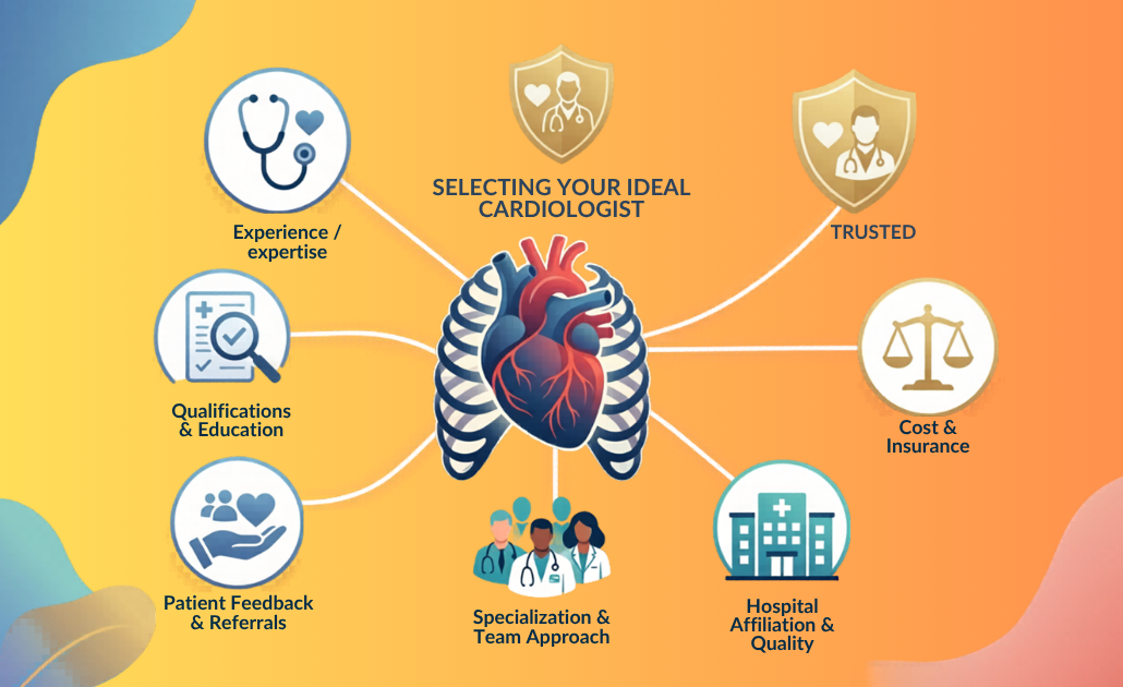
When subtle warning signs emerge, such as sudden breathlessness, unexplainable exhaustion, or sharp chest twinges, finding a qualified heart doctor in Lucknow is vital. However, for residents in Uttar Pradesh, filtering through endless medical directories to find an elite heart doctor in Lucknow can quickly become an overwhelming task.

Choosing who protects your cardiovascular system shouldn’t be left to random online searches or luck. To find a dependable heart doctor who can guide you toward recovery, you must know what specific qualities, medical qualifications, and clinical facilities to look for. To help you make an informed, confident decision, we have distilled the selection process down to five non-negotiable criteria to locate the ideal heart doctor in Lucknow.

1. Verify Academic Credentials and Specialization

Not all heart conditions are treated equally, and not all medical specialists perform the same role. Cardiology is divided into highly specific sub-specialties. When searching for a heart doctor, it is crucial to understand the distinct qualifications of the medical professional you are considering:

  • Cardiologist (Non-Invasive/Clinical): Focuses on the prevention, diagnosis, and management of heart disease through lifestyle changes and medications. A clinical heart doctor in Lucknow manages hypertension, cholesterol, and initial evaluations like ECGs or Echo scans.
  • Interventional Cardiologist: Specialized in performing advanced, minimally invasive catheter-based procedures. If you require a coronary angioplasty, stent placement, or structural repairs like TAVI (Transcatheter Aortic Valve Implantation), you need an experienced interventional heart doctor in Lucknow.
  • Cardiothoracic Surgeon: The surgical specialist who steps in when structural blockages or defects require complex open-heart interventions, such as a Coronary Artery Bypass Graft (CABG) or valve replacements.

Look for core qualifications such as an MD followed by a DM in Cardiology or an M.Ch for surgical branches from recognized premium medical institutions when vetting a heart doctor in Lucknow.

2. Evaluate Clinical Experience and Procedure Volume

Medical knowledge is vital, but hands-on experience builds clinical precision. When evaluating a heart doctor in Lucknow, consider their years in active practice and their procedural track record.

  • Consider their clinical background:
  • How many years have they practiced as a core heart doctor in Lucknow?
  • If you need an invasive intervention, how frequently does that specific heart doctor in Lucknow perform that procedure?

A high volume of successful procedures typically translates to smoother execution, a lower risk of clinical complications, and more seasoned decision-making when handling complex or unexpected situations.

3. Hospital Infrastructure and Emergency Readiness

A heart doctor in Lucknow is only as effective as the environment they operate in. Cardiac emergencies—like an acute heart attack—demand split-second responses where every single minute dictates the preservation of heart muscle tissue.

When choosing a specialist, evaluate the infrastructure of the hospital or clinic where your chosen heart doctor in Lucknow practices:

  • The Cath Lab: Is there a fully operational, 24/7 Digital Cardiac Catheterization Lab equipped to handle emergencies immediately?
  • Support System: Does the facility house a dedicated Cardiac Intensive Care Unit (CICU) staffed by trained cardiac nurses?
  • Multidisciplinary Care: Can the hospital seamlessly bring together clinical cardiologists, electrophysiologists, and surgeons under one roof for complex cases?

Opting for a heart doctor in Lucknow affiliated with a well-equipped, responsive healthcare ecosystem ensures that you are protected both during routine checks and sudden crises.

Comprehensive medical infographic breaking down clinical qualifications, advanced facility standards, cardiac red flags, and patient partnership metrics for choosing a top cardiologist.

4. Prioritize a Patient-Centered Communication Style

Cardiovascular disease is frequently a long-term journey rather than a one-time fix. Managing it demands lifestyle overhauls, consistent medication, and ongoing medical checkups. This means your relationship with your heart doctor in Lucknow will likely span years.

During your initial consultation, assess how the doctor communicates:

  • Does the doctor take the time to explain your diagnostic scans and blood panels in plain language?
  • Are they willing to map out treatment pathways and discuss the pros and cons of medications versus procedures?
  • Do they listen patiently to your lifestyle concerns, or do they rush through the appointment?

A great heart doctor in Lucknow treats the person, not just the condition. Empathy and clear communication improve patient confidence and reduce stress.

5. Assess Accessibility and Emergency Support

Heart issues don’t stick to a convenient 9-to-5 schedule. Beyond standard clinic timings, consider how accessible your is during off-hours:

  • Location Convenience: Is the clinic or hospital easily accessible from your residence to minimize travel strain?
  • Emergency Protocol: What is the specific protocol if you experience sudden symptoms at night? Is there an emergency contact helpline or an on-call team coordinated by your heart doctor in Lucknow?
  • Teleconsultation Options: Does the doctor provide virtual consults for minor adjustments to medications or routine follow-up questions?

A Lifesaving Rule of Thumb

Never delay a cardiac checkup out of fear or inconvenience. If you are over the age of 40, possess a family history of heart conditions, or manage chronic health markers like diabetes or high blood pressure, regular diagnostic screenings with a trusted heart doctor in Lucknow are your best defense.

Taking the time to evaluate a specialist based on these foundational pillars gives you total control over your health roadmap. Your heart deserves nothing less than meticulous, highly qualified, and deeply compassionate care from the finest heart doctor in Lucknow.

About the Author

Dr. Gautam Swaroop

Sr. Interventional Cardiologist

Harispandan Heart Care & Sleep Clinic – Best Heart Care Clinic Gomti Nagar, Lucknow

Dr. Gautam Swaroop is an experienced interventional cardiologist specializing in advanced heart procedures like TAVI/TAVR, MitraClip, complex angioplasties, and structural heart disease management. He is passionate about patient education and preventive cardiology, making complex heart health topics simple and easy to understand.

FAQs

1. When should I see a heart doctor in Lucknow?

See a specialist immediately if you experience chest pain, unexplained breathlessness, dizziness, or chronic palpitations. Proactive screening is highly recommended if you are over 40 with a family history of heart disease, diabetes, or high blood pressure.

2. Clinical vs. Interventional Cardiologist: What’s the difference?

A clinical cardiologist manages heart conditions using medications and lifestyle changes. An interventional heart doctor in Lucknow performs advanced, minimally invasive procedures inside a Cath Lab, such as angioplasty and stent placement.

3. What should I bring to my first appointment?

Bring all recent blood work (lipid/sugar profiles), past cardiac reports (ECG, Echo, TMT), a complete list of your current medications with exact dosages, and a record of your family medical history.

4. Why does the hospital’s infrastructure matter?

Cardiac emergencies require immediate action. The affiliated hospital must have a 24/7 operational Cath Lab, a dedicated Cardiac ICU (CICU), and an on-call surgical team to handle sudden complications without delay.

5. Can heart disease be cured without surgery?

Yes. Many early-stage conditions can be managed effectively without surgery. A combination of targeted medical therapy, a heart-healthy diet, regular physical exercise, and stress management is often highly successful.