
ECG (Electrocardiogram): Purpose, Procedure, Results, Benefits & When You Need One
Quick Answer: What is an ECG?
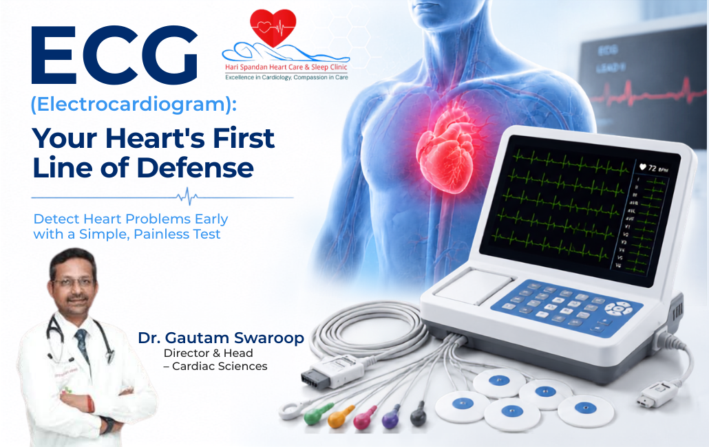
An ECG, or Electrocardiogram, is a quick, painless, and non-invasive medical test. Specifically, it measures and records the electrical activity of your heart. Primarily, doctors use it to evaluate your heart rhythm and detect arrhythmias. Furthermore, it helps identify signs of a heart attack. Consequently, it serves as one of the most vital frontline cardiac diagnostic tests.
Introduction
Currently, cardiac issues are rising among young adults. Additionally, India faces a high prevalence of diabetes and hypertension. Therefore, being proactive about heart health is more critical than ever. Recently, you might have experienced chest pain, a racing heartbeat, or sudden breathlessness. In that case, your doctor will likely recommend an ECG.
Medical professionals also call this test an electrocardiogram. Indeed, this simple yet powerful test remains a cornerstone of modern cardiology. Furthermore, diagnostic centres across India include it standardly in “Master Health Check-up” packages. But what exactly happens during this test? Moreover, what can the results tell you about your heart?
As a cardiologist, I often explain a simple concept to my patients. Namely, the heart operates on its own internal electrical wiring. Specifically, an electrical impulse triggers every single heartbeat. Meanwhile, an ECG machine acts like a highly sensitive antenna. It picks up these signals and translates them into a readable graph.
Perhaps you are preparing for a routine health screening. Alternatively, you might be managing a chronic condition or seeking an emergency chest pain evaluation. Regardless, understanding your heart’s electrical activity is your first line of defence. Ultimately, this comprehensive guide will walk you through the ECG procedure. Next, it will explain the results and highlight the importance of this preventive test.
Table of Contents
- What Is an ECG?
- How Does an ECG Work?
- Why Is an ECG Done?
- What Conditions Can an ECG Detect?
- Types of ECG Tests
- How Is an ECG Performed?
- Understanding ECG Results
- Benefits of an ECG
- Risks and Limitations of an ECG
- Who Should Get an ECG?
- How to Prepare for an ECG
- What Happens After an ECG?
- Frequently Asked Questions
What Is an ECG?
Medical Definition Box:
An ECG (Electrocardiogram) is a diagnostic medical tool. Doctors use it to evaluate the electrical impulses generated by the heart. First, technicians place small sensors (electrodes) on your skin. Then, the test records the timing and strength of these signals. Ultimately, it tracks them traveling through the heart muscle.
Furthermore, we cannot overstate the medical significance of an ECG. Actually, it is the absolute gold standard for rapid evaluations. Specifically, it helps patients presenting with heart-related symptoms in any clinic or emergency room. Because damaged or stressed heart muscle alters electrical patterns, an ECG report provides immediate, life-saving insights.
Therefore, cardiologists highly recommend an ECG. Ultimately, it gives a real-time “snapshot” of your heart’s health. In the world of heart disease diagnosis, time is muscle. Consequently, a faster tracing allows us to diagnose critical issues like a heart attack quickly. Then, we can initiate life-saving treatment.
What Does ECG Stand For?
- Full Form: Electrocardiogram.
- Pronunciation: ih-lek-troh-KAHR-dee-uh-gram.
- Medical Meaning: Let us break the word down. First, “Electro” refers to electrical activity. Second, “Cardio” refers to the heart. Finally, “Gram” means a record or picture. Simply put, it is a recorded picture of the heart’s electrical activity.
- Why It Is Searched: Many people search for the “ECG meaning” online. Specifically, doctors and lab technicians use the abbreviation universally. However, busy clinics rarely explain the actual test mechanics to patients.
How Does an ECG Work?
Initially, your heart relies on a natural pacemaker called the sinus node. This node generates an electrical spark that travels through the heart muscle. Consequently, this spark causes the heart to squeeze and pump blood throughout your body.
During an electrocardiogram test, technicians place small, sticky patches called electrodes on your chest, arms, and legs. Next, they connect these electrodes to an ECG machine using wires. Importantly, the machine does not send any electricity into your body. Instead, it simply listens to and amplifies your heart’s tiny natural electrical signals.
Subsequently, the machine prints these signals on special grid paper or displays them on a digital screen as waveforms. Generally, doctors analyse three main parts of this wave:
- The P Wave: This represents the upper chambers of the heart (atria) squeezing to push blood down.
- The QRS Complex: This represents the lower, larger chambers (ventricles) squeezing to pump blood out to the rest of the body.
- The T Wave: Finally, this represents the heart resetting and relaxing before the next heartbeat.
What Does an ECG Measure?
By analysing the size, shape, and timing of these waves, a cardiologist can measure several things:
- Heart Rate: How fast your heart is beating, detecting abnormally fast or slow rates.
- Heart Rhythm: Whether the beat is steady, skipped, or irregular.
- Electrical Conduction: How smoothly the electrical signal travels through the heart tissue.
- Signs of Heart Muscle Damage: Whether certain areas of the heart lack oxygen.
- Heart Enlargement: Whether the walls of the heart are abnormally thick, often caused by unmanaged high blood pressure.
Why Is an ECG Done?
Typically, a doctor or cardiologist will order an ECG to evaluate a variety of symptoms and situations. Specifically, the most common reasons include:
- Chest pain or discomfort: To rule out or confirm an active heart attack.
- Palpitations: To investigate a fluttering, racing, or pounding heartbeat.
- Shortness of breath: To see if a weakened heart is causing breathing difficulties.
- Dizziness or Lightheadedness: To check for a slow or irregular heart rhythm affecting blood flow to the brain.
- Fainting (Syncope): To identify potentially dangerous electrical blocks in the heart.
- High blood pressure (Hypertension): To check if the heart has become enlarged due to stress.
- Irregular heartbeat (Arrhythmia): To map out the exact type of irregularity.
- Routine master health check-ups: As part of a preventive annual screening.
- Pre-operative evaluation: To ensure your heart is strong enough to handle surgery and anaesthesia.
What Conditions Can an ECG Detect?
Indeed, an ECG is a highly versatile cardiac diagnostic test. The table below outlines the primary conditions an ECG can detect. Furthermore, it details what the doctor looks for on the printed report.
| Medical Condition | Description | Common ECG Findings |
|---|---|---|
| Heart Attack (Myocardial Infarction) | A blockage stops blood flow to the heart muscle, causing tissue death. | ST-segment elevation or depression, deep Q waves. |
| Previous Heart Attack | Scar tissue remains from a past, sometimes unnoticed, silent heart attack. | Abnormal Q waves indicate dead heart muscle. |
| Arrhythmias | The heart beats too fast, too slow, or irregularly (e.g., Atrial Fibrillation). | The report shows irregular distances between the QRS complexes or absent P waves. |
| Heart Block | The electrical signal delays or stops as it travels through the heart. | Prolonged PR intervals or dropped QRS beats appear. |
| Enlarged Heart (Hypertrophy) | The heart walls thicken, often due to high blood pressure. | Unusually tall QRS waves indicate extra muscle mass. |
| Coronary Artery Disease | Narrowed arteries reduce blood flow (Ischemia). | Doctors spot T-wave inversions or ST depressions. |
| Cardiomyopathy | A heart muscle disease makes pumping blood harder. | Low voltage waves and bundle branch blocks show up. |
| Electrolyte Imbalance | Blood contains abnormal levels of potassium or calcium. | Peaked T waves indicate high potassium, or T waves flatten. |
Types of ECG Tests
However, not all heart symptoms happen while you sit in a clinic. Therefore, doctors use different types of electrocardiogram tests. Consequently, they can capture heart activity in various states.
| Type of ECG Test | Purpose & Description | Duration | Best Used For |
|---|---|---|---|
| Resting ECG | Measures heart activity while you lie down comfortably. | 5–10 minutes. | Routine check-ups and resting chest pain evaluation. |
| Stress ECG (Treadmill Test / TMT) | Measures heart activity while you walk on a treadmill. | 15–30 minutes. | Diagnosing blocked arteries that only cause symptoms during physical exertion. |
| Holter Monitor | A portable ECG worn continuously to record all heartbeats. | 24–48 hours. | Catching unpredictable palpitations or silent arrhythmias as you go about your day. |
| Event Monitor | A portable device you activate only when you feel symptoms. | Up to 30 days. | Diagnosing rare symptoms like occasional fainting or a suddenly racing heart. |
(Internal Link Opportunity: Learn more about our Treadmill Test (TMT) and Holter Monitoring services).
How Is an ECG Performed?
Overall, the ECG procedure is remarkably straightforward. Furthermore, it requires minimal effort from the patient. Here is the step-by-step process of what you should expect at a clinic:
- Patient Preparation: First, the staff will ask you to lie flat and comfortably on an examination couch. Next, you will need to expose your chest, wrists, and ankles.
- Skin Preparation: Then, the technician will wipe your skin with a spirit swab. If you have a hairy chest, they may shave small areas. Consequently, this ensures the sticky patches adhere properly and record a clear signal.
- Electrode Placement: Next, the technician will attach 10 to 12 sticky patches (electrodes) to specific locations on your chest, arms, and legs.
- Recording Heart Signals: The technician connects the electrodes to the ECG machine via cables. Subsequently, you must lie very still, not talk, and breathe normally. Meanwhile, the machine records your heart’s electrical activity.
- Test Duration: The actual recording takes only a few seconds. Ultimately, the entire appointment usually lasts less than 10 minutes.
- Removing Electrodes: Once the recording is complete, the technician will peel off the electrodes. Then, you can immediately wipe off any residual gel and put your clothes back on.
- Report Interpretation: Finally, the machine prints out the ECG report instantly. Afterward, your cardiologist will review it.
Is an ECG Painful?
No. An ECG is completely painless.
Specifically, it is a strictly non-invasive test. The machine only reads your heart’s electrical signals. Therefore, it does not send any electrical current into your body. Consequently, you will not feel any shocks or vibrations. Removing the sticky patches at the end may feel like pulling off a mild adhesive bandage. However, the procedure is entirely safe for adults, children, and pregnant women.
Understanding ECG Results
Once you finish the test, your doctor will classify your results as either a Normal ECG or an Abnormal ECG.
| Result Type | What It Means | Next Steps |
|---|---|---|
| Normal ECG | Your heart rate falls between 60-100 beats per minute, and the rhythm is steady. Furthermore, the electrical waves look healthy. Consequently, they indicate no current signs of damage or strain. | You require no further immediate testing unless your symptoms persist. |
| Abnormal ECG | The test shows irregularities in heart rate, rhythm, or waveform shape. This could indicate a previous heart attack, current lack of oxygen, enlarged heart, or arrhythmia. | Your cardiologist will likely order additional tests. For example, they might order a 2D Echo or blood tests like a Troponin test. |
Disclaimer: An abnormal result does not always mean you have severe heart disease. Sometimes, natural variations, anxiety, or harmless irregularities can trigger an abnormal reading. Therefore, only a qualified medical professional should interpret an ECG.
Benefits of an ECG
Why is the ECG the most commonly performed cardiology test globally and across India? Primarily, it offers unparalleled advantages for patient care:
- Quick & Immediate: The machine prints results instantly. Thus, doctors can make life-saving split-second decisions during a heart attack.
- Completely Safe: It uses no radiation (unlike X-rays), requires no injections, and has no side effects.
- Highly Affordable: It is one of the most cost-effective medical tests available in Indian healthcare.
- Non-Invasive: It requires no needles or surgical incisions.
- Early Detection: It can catch heart conditions before they cause severe, irreversible symptoms.
- Guides Treatment: It helps doctors decide if you need daily medication, a pacemaker, or an emergency angioplasty.
- Widely Available: You can find it in almost all primary health centres, clinics, and diagnostic labs.
Risks and Limitations of an ECG
While an ECG is a phenomenal diagnostic tool, it does have a few limitations. Therefore, patients should understand the following:
- It is a snapshot in time: An ECG only shows what your heart is doing at the exact moment of the test. For example, if your abnormal heartbeat comes and goes, a 10-second resting ECG might miss it completely.
- Cannot detect all blockages: An ECG cannot directly see inside your coronary arteries. You could have a blocked artery. However, if it does not restrict blood flow enough to stress the resting heart muscle, the ECG may look completely normal (a “false-negative”).
- Temporary abnormalities: Sometimes, anxiety, shivering, or poor electrode contact can create a messy reading. Doctors call this an “artefact”. Consequently, the technician must repeat the test.
- Need for further testing: An abnormal ECG often requires confirmation. Specifically, doctors might order an Echocardiogram (ultrasound of the heart) or a Coronary Angiogram to get a complete, accurate picture.
Who Should Get an ECG?
Research shows that Indians are genetically predisposed to cardiovascular diseases almost a decade earlier than Western populations. Therefore, preventive heart screening is vital. You should strongly consider getting an ECG if you fall into any of the following categories:
- Adults Over 35-40: Aging naturally increases the risk of heart disease. Consequently, doctors highly recommend a baseline ECG.
- Diabetics: High blood sugar damages blood vessels over time, silently harming the heart.
- Hypertensive Patients: High blood pressure forces the heart to work harder, leading to muscle thickening.
- High Cholesterol Patients: Plaque buildup can restrict blood flow, altering the heart’s electrical signals.
- Family History: Genetics play a massive role in premature heart attacks and blockages.
- Smokers / Tobacco Users: Tobacco use dramatically increases the risk of coronary artery disease.
- Anyone with Symptoms: Chest pain, breathlessness on exertion, or sudden dizziness require immediate ECG evaluation.
How to Prepare for an ECG
Preparing for an ECG is incredibly simple. Therefore, follow these patient-focused guidelines to ensure an accurate reading:
- Clothing: Wear a loose shirt, kurta, or a top that you can easily unbutton or remove. Additionally, avoid restrictive clothing or full-body dresses.
- Skin Preparation: Do not apply body lotions, heavy oils, or talcum powder to your chest or limbs on the day of the test. Otherwise, they can prevent the electrodes from sticking properly.
- Food and Drink: You do not need to fast. Furthermore, you can eat your regular meals and drink water normally. However, avoid heavy caffeine (strong coffee/tea) or smoking right before the test. Consequently, these can artificially raise your heart rate.
- Medication: Continue taking all your prescribed medications. Do this unless your doctor explicitly tells you to pause them.
- Before the Test: Relax. First, sit quietly in the waiting area for a few minutes. Rushing up the stairs or feeling anxious can temporarily alter your resting heart rhythm.
What Happens After an ECG?
Because the test results are immediate, you won’t have to wait days for a lab to process them.
- Receiving Results: The ECG machine prints a graph right in the examination room.
- Cardiologist Review: Your doctor will review the waveforms. Specifically, they will look for patterns that suggest damage, blockages, or electrical misfires.
- Treatment Planning: If the ECG is normal and your symptoms are mild, you may go home with peace of mind. However, if there is a minor abnormality, your doctor may prescribe medications. These will help manage your heart rhythm or control blood pressure.
- Additional Tests: If the ECG suggests a more serious issue, your doctor may immediately refer you for an Echocardiography (2D Echo) or a Treadmill Test (TMT). Consequently, these tests evaluate your heart’s function under stress.
Frequently Asked Questions
1. What is an ECG?
An ECG (Electrocardiogram) is a non-invasive diagnostic test that records the electrical activity of the heart. Specifically, it uses small sensors placed on the skin to detect electrical signals. Then, it graphs these signals on paper. Consequently, it helps doctors quickly evaluate heart rate, rhythm, and signs of muscle damage.
2. What does ECG stand for?
ECG stands for Electrocardiogram. First, “Electro” refers to the electrical impulses of the heart. Second, “cardio” refers to the heart itself. Finally, “gram” means a visual record or tracing.
3. Why is an ECG done?
Doctors do an ECG to investigate heart-related symptoms like chest pain, shortness of breath, palpitations, and dizziness. Furthermore, they routinely use it for preventive master health check-ups. Additionally, it monitors the effectiveness of prescribed heart medications.
4. Is an ECG painful?
No, an ECG is completely painless. Indeed, it is a non-invasive procedure that does not send any electricity into the body. The machine merely passively records the natural electrical impulses of your heart.
5. How long does an ECG take?
The actual recording of the heart’s electrical signals takes only a few seconds. However, the entire process usually takes about 5 to 10 minutes in total. This includes undressing, placing the electrodes on your chest and limbs, and removing them afterward.
6. Can an ECG detect a heart attack?
Yes. An ECG is the primary tool used to diagnose an acute heart attack (myocardial infarction). When a blocked artery deprives the heart muscle of oxygen, its electrical patterns change drastically. Consequently, this is immediately visible on the ECG graph.
7. Can an ECG detect blocked arteries?
An ECG cannot directly visualize blocked arteries. However, it can detect the effects of a blocked artery. It does this if the blockage is severe enough to deprive the resting heart of oxygen. If doctors suspect blockages but the resting ECG is normal, they usually recommend a Treadmill Test (TMT).
8. Is fasting required before an ECG?
No, fasting is not required before a standard resting ECG. Therefore, you can eat and drink normally before your appointment. However, it is advisable to avoid consuming strong tea, coffee, or energy drinks just before the test.
9. What is the difference between ECG and Echo?
An ECG records the electrical activity of the heart, identifying rhythm issues and electrical blocks. Conversely, an Echocardiogram (2D Echo) is an ultrasound that looks at the mechanical structure of the heart. It evaluates how well the heart valves are working and how strong the heart muscle is pumping.
10. How often should I get an ECG?
If you are over 40 or have risk factors like diabetes, hypertension, or a family history of heart disease, you should get an annual ECG during your routine health check-up. If you have an existing heart condition, your cardiologist will advise on the frequency.
11. Is an ECG safe during pregnancy?
Yes, an ECG is entirely safe during pregnancy. Because it is non-invasive and does not involve radiation (unlike X-rays) or electrical shocks, it poses absolutely no risk to the mother or the developing baby.
12. What do abnormal ECG results mean?
An abnormal ECG result means the heart’s electrical pattern deviated from the standard rhythm. This could indicate an arrhythmia, an enlarged heart, an electrolyte imbalance, or a lack of blood flow. Therefore, an abnormal result always requires a doctor’s review.
13. Which doctor performs an ECG?
A general physician, an emergency room doctor, or a specialist can order an ECG. Typically, a trained nurse or lab technician administers the test. However, a Cardiologist best interprets the complex results.
14. How much does an ECG cost in India?
The cost of an ECG in India is very affordable. Typically, it ranges between ₹200 to ₹800 depending on the city, the type of clinic, or the hospital. Overall, it is a highly cost-effective diagnostic tool that provides crucial information about your heart health.
15. When should I get an ECG immediately?
You should seek emergency medical care and get an immediate ECG if you experience sudden, severe chest pain (especially if it radiates to the left arm, jaw, or back). Furthermore, you need one if you experience extreme shortness of breath, unexplained fainting, or heavy sweating combined with a pounding heart.
Key Takeaways
- An ECG (Electrocardiogram) is a fast, safe, and non-invasive test that records the electrical activity of your heart.
- It is the primary, frontline test used to evaluate chest pain. Furthermore, it can immediately detect a heart attack or abnormal rhythms (arrhythmias).
- The test takes less than 10 minutes, involves placing sticky electrodes on your chest and limbs, and is completely painless.
- An ECG cannot detect everything. While it is excellent for electrical issues, hidden blockages may require a Treadmill Test (TMT) or an Echocardiogram.
- Given the rising heart health issues in India, adults over 40, diabetics, smokers, and individuals with high blood pressure should get routine preventive heart screenings, including an ECG.
Conclusion
Understanding your heart’s health doesn’t have to be complicated. Indeed, the ECG remains one of the most brilliant and fundamental tools in modern cardiology. Specifically, it translates your heart’s invisible electrical impulses into a readable graph. Consequently, an electrocardiogram provides doctors with immediate, life-saving information.
Perhaps you are dealing with unexplained palpitations. Alternatively, you might be managing diabetes and hypertension. Or, you simply want to stay proactive about your health as you age. In any case, getting a timely ECG is a vital step toward long-term cardiovascular wellness. Therefore, do not wait for severe symptoms to appear. Prioritize your heart health through early diagnosis and regular preventive screenings.
Book your Appointment now
Are you experiencing chest pain, palpitations, dizziness, or shortness of breath? Do not ignore what your heart might be telling you. Therefore, schedule a comprehensive cardiology consultation and ECG evaluation at Harispandan Clinic today. Ultimately, our expert cardiac team is dedicated to providing you with accurate diagnoses, personalized care, and peace of mind. Book your appointment now and take the first step toward a healthier heart!
Medical Disclaimer: This article is intended for educational and informational purposes only and does not replace professional medical advice, diagnosis, or treatment. Always seek the advice of a qualified healthcare provider with any questions you may have regarding a medical condition. If you are experiencing symptoms of a heart attack, severe chest pain, or a medical emergency, seek emergency medical care immediately.